
Knee osteoarthritis is one of the most prevalent joint disorders in Egypt, affecting over 30% of adults aged 50 and above. It is directly associated with elevated body weight, repeated deep squatting, and uncorrected genu varum (bowed legs). Osteoarthritis is a progressive, irreversible degenerative process—damaged hyaline cartilage does not regenerate—yet disease progression and clinical symptoms can be substantially controlled, particularly during the early stages. This guide outlines the anatomical grades, evidence-based management strategies, and practical methods to protect the joint from accelerating deterioration.
What is Knee Osteoarthritis?
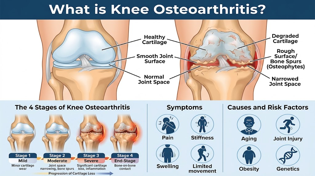
Knee osteoarthritis is the progressive degeneration of the articular (hyaline) cartilage that lines the distal femur, proximal tibia, and the posterior surface of the patella. Healthy articular cartilage measures 3 to 5 mm in thickness with a smooth, low-friction surface facilitating near-frictionless gliding. As degeneration sets in, the cartilage thins, fibrillates, and fissures until the subchondral bone is exposed, resulting in bone-on-bone friction, mechanical pain, crepitus, stiffness, and reactive effusion.
The disease is classified radiographically into four grades using the Kellgren-Lawrence grading system:
- Grade 1: Minimal (doubtful joint space narrowing and possible osteophytic lipping).
- Grade 2: Mild (definite osteophytes with possible mild joint space narrowing).
- Grade 3: Moderate (multiple moderate osteophytes, definite joint space narrowing, and some subchondral sclerosis).
- Grade 4: Severe (marked joint space narrowing, extensive subchondral sclerosis, and large osteophytes—bone-on-bone contact).
Progression across these grades can take several years and is heavily influenced by biomechanical loading, physical activity levels, and early intervention.
Causes and Risk Factors of Knee Osteoarthritis
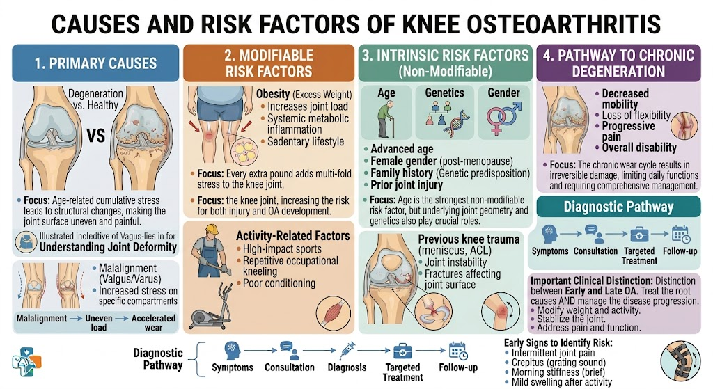
The pathophysiology involves multifactorial mechanical, systemic, and metabolic drivers:
- Age Over 50: The most prevalent non-modifiable factor, as chondrocytes lose reparative capacity and the extracellular matrix loses elasticity.
- Obesity: The single most important modifiable risk factor. In Egypt, adult obesity prevalence exceeds 35%. Every additional kilogram of body weight increases the compressive load on the tibiofemoral joint by 3 to 5 kg per step.
- Deep Squatting and Floor Sitting: Full knee flexion drives patellofemoral compressive stresses up to 7 times body weight—a common daily postural habit that accelerates chondral breakdown.
- Genu Varum (Bowlegged Deformity): Concentrates 60–80% of total mechanical axial load through the medial (inner) compartment rather than an even 50%, accelerating inner compartment wear 2- to 4-fold.
- Previous Intra-Articular Injuries: An untreated anterior cruciate ligament (ACL) rupture creates chronic micro-instability that accelerates degenerative changes. Similarly, prior meniscectomy removes vital shock absorption, shifting mechanical load directly onto the articular surface.
- Type 2 Diabetes Mellitus: Affects over 15% of adults in Egypt; chronic hyperglycemia induces advanced glycation end-products (AGEs) within the collagen framework, compromising cartilage elasticity.
- Quadriceps Weakness: The quadriceps musculature serves as the primary dynamic shock absorber for the knee; insufficient muscle tone transfers repetitive impacts straight into the articular cartilage.
Symptoms of Knee Osteoarthritis
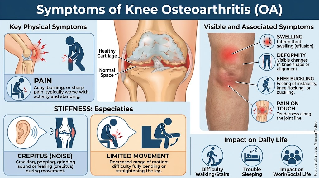
Clinical manifestations correspond to the anatomical severity of cartilage wear:
- Early Stages: Pain initiated upon climbing stairs, descending slopes, or rising from a seated position (start-up pain) that eases with gentle mobilization; morning stiffness lasting under 30 minutes; joint crepitation on passive or active flexion.
- Advanced Stages: Persistent pain even at rest or during the night, recurrent inflammatory effusions, restricted range of motion, antalgic gait (limping), and progressive angular deformity of the leg.
Core Clinical Signs:
- Pain on Navigating Stairs: Frequently the initial presentation, reflecting amplified patellofemoral and medial compartment loads.
- Morning Stiffness: Degenerated surfaces lack fluid lubrication after prolonged rest; typically resolves within 15 to 30 minutes of light activity (stiffness exceeding 30–60 minutes suggests an underlying systemic inflammatory arthropathy such as rheumatoid arthritis).
- Audible or Palpable Crepitus: A rough, grating sensation during knee flexion caused by uneven, fibrillated articular surfaces.
- Recurrent Joint Effusion: Fluid accumulation following weight-bearing stress, indicating secondary mechanical synovitis.
- Progressive Varus Malalignment: In late stages, medial compartment cartilage loss narrows the medial joint space, gradually increasing the bowlegged appearance.
Can One Live with Knee Osteoarthritis?
Adaptation depends on the clinical grade:
- Grades 1–2: Highly manageable. A combined regimen of weight loss (5–10 kg), targeted quadriceps strengthening, minimizing deep squats, avoiding excessive stair climbing, and utilizing intermittent NSAIDs during flare-ups allows patients to remain active for years.
- Grade 3: Manageable with intensive physical therapy, offloading braces, and targeted intra-articular therapy, though structural progression persists.
- Grade 4: Difficult to tolerate. Unremitting mechanical pain compromises ambulation, functional independence, and restorative sleep; joint arthroplasty represents the definitive option.
Many patients maintain functional independence across Grades 2 and 3 through lifestyle modifications: utilizing chairs instead of sitting on the floor, using elevators when available, praying from a chair during severe flares, and maintaining a healthy body weight. The clinical goal is decelerating joint deterioration rather than expecting cartilage regeneration.
Prevention of Knee Osteoarthritis
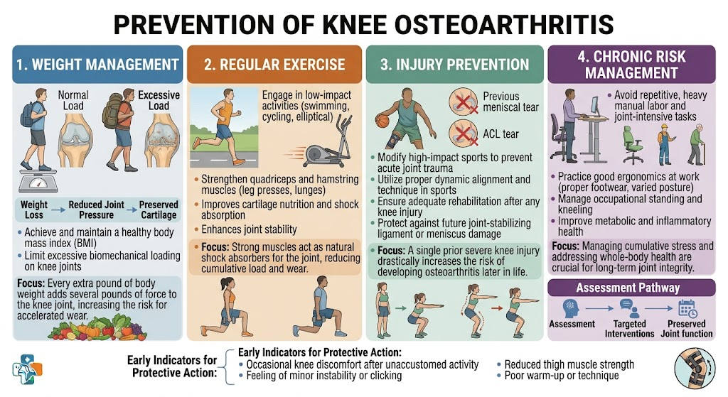
Preventive strategies should be established prior to the onset of irreversibility:
- Weight Management: Shedding 5 kg decreases compressive knee loads by 15 to 25 kg per step, cutting the long-term risk of symptomatic osteoarthritis by up to 50%.
- Quadriceps Conditioning: Engaging in daily targeted exercise (e.g., straight leg raises, 3 sets of 15 repetitions) builds dynamic stabilization and dissipates impact forces away from articular surfaces.
- Limiting Deep Hyperflexion: Utilizing low stools for domestic chores rather than full squats relieves up to 70% of patellofemoral pressure.
- Prompt Treatment of Traumatic Injuries: Untreated ACL ruptures induce osteoarthritis within 5 to 10 years. Preserving and repairing meniscal tissue via arthroscopic repair is far superior to meniscectomy whenever feasible.
- Early Realignment Osteotomy: Correcting severe varus malalignment in young, active patients through high tibial osteotomy unloads the damaged medial compartment, delaying total joint replacement by 10 to 15 years.
- Low-Impact Physical Activity: Swimming, stationary cycling, and walking on level surfaces sustain synovial fluid circulation and chondrocyte nutrition without high mechanical impact.
Treatment of Knee Osteoarthritis
Management follows a graded approach based on disease stage:
| Grade | Primary Treatment | Adjunctive Measures | Surgical Intervention (If Indicated) |
| 1 (Early softening/fibrillation) | Weight loss + Quadriceps strengthening + Activity modification | Short-course NSAIDs during flare-ups | Rarely indicated |
| 2 (Superficial fissuring) | Structured physical therapy + Low-impact conditioning | Viscosupplementation (HA) or PRP injections | Diagnostic/therapeutic arthroscopy in select mechanical pathologies |
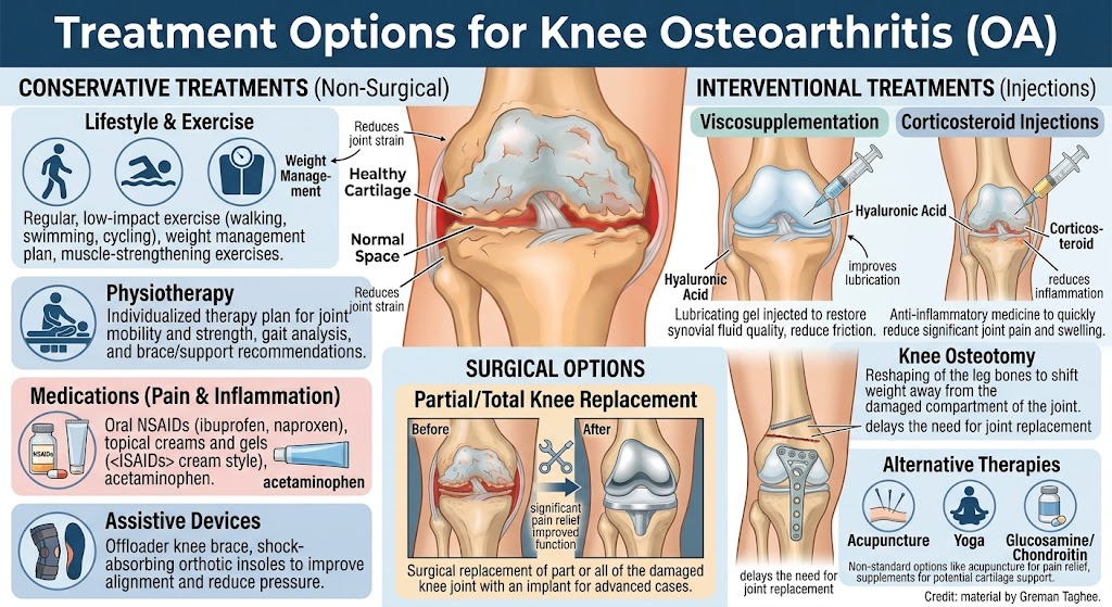
| 3 (Deep fissuring, osteophytes) | Intensive rehabilitation + Offloader unloader brace + Orthotics | Repeated biologic/lubricant injections | Corrective osteotomy (isolated medial disease) or partial (unicompartmental) knee replacement |
| 4 (Bone-on-bone contact) | Analgesics + Activity pacing + Walking aids | Intra-articular injections for temporary symptomatic relief | Total Knee Arthroplasty (TKA) |
Weight loss represents the most cost-effective non-pharmacological treatment: a 10 kg reduction yields symptom improvement comparable to systemic analgesics, with enduring cardiovascular and systemic advantages. Strong quadriceps absorb 30–40% of ambulatory shock waves, substantially reducing direct stress on subchondral bone.
Intra-articular interventions (hyaluronic acid or platelet-rich plasma) can reduce pain and improve joint kinematics for 6 to 12 months, though they do not re-grow lost cartilage. For Grade 4 disease, Total Knee Arthroplasty (TKA) serves as the definitive solution—alleviating pain in 90–95% of patients with an expected implant survivorship of 15 to 25 years.
Frequently Asked Questions About Knee Osteoarthritis
Evidence-based answers to etiology, staging, diagnostic radiographic parameters, medical preservation, and modern surgical options.
What is Knee Osteoarthritis (Gonarthrosis)?
Knee osteoarthritis is a progressive degenerative disorder involving the entire synovial joint organ. While pathologically identified by the gradual breakdown of articular hyaline cartilage, it encompasses subchondral bone sclerosis, osteophytosis (bone spur development), synovial lining hyperplasia, meniscus extrusion, and ligamentous laxity that culminate in joint dysfunction and pain.
What are the hallmark symptoms of knee osteoarthritis?
Typical clinical findings include:
• Mechanical Activity-Related Pain: A dull, deep ache that exacerbates during weight-bearing activities (especially stair ascent/descent and prolonged standing) and improves with rest.
• Gelling / Morning Stiffness: Brief stiffness lasting fewer than 30 minutes following periods of inactivity.
• Crepitus & Decreased Arc of Motion: Palpable or audible grinding sensations during knee flexion-extension, progressive loss of terminal extension, and joint line tenderness.
What is the difference between primary and secondary knee osteoarthritis?
Primary osteoarthritis develops insidiously without a single triggering event, predominantly driven by intrinsic cellular senescence, genetic predisposition, female sex, and biomechanical wear over time.
Secondary osteoarthritis stems from an identifiable underlying cause—most commonly post-traumatic intra-articular fractures, prior meniscectomy, chronic anterior cruciate ligament (ACL) insufficiency, congenital/developmental skeletal dysplasias, or previous septic arthritis.
How is knee osteoarthritis diagnosed and graded by orthopedic specialists?
Diagnosis is confirmed using standardized weight-bearing (standing) radiographs. The Kellgren-Lawrence (KL) grading system classifies disease severity:
• Grade 1 (Doubtful): Minute osteophytic lipping with doubtful joint space narrowing.
• Grade 2 (Mild): Definite osteophytes with possible joint space narrowing (unimpaired joint architecture).
• Grade 3 (Moderate): Definite joint space narrowing, moderate multiple osteophytes, and early subchondral sclerosis.
• Grade 4 (Severe): Marked joint space obliteration (bone-on-bone contact), severe subchondral sclerosis, and notable bony deformity.
Why is weight management critical in treating knee osteoarthritis?
Biomechanical studies demonstrate that each 1 kg of body mass lost reduces joint load by approximately 4 kg with each foot strike during walking. Beyond mechanical unloading, adipose tissue functions as an active endocrine organ secreting systemic pro-inflammatory cytokines and adipokines (such as leptin and resistin). Achieving a 5% to 10% weight loss blunts these inflammatory cascades and significantly alleviates pain.
What role does physical therapy play in conservative management?
Physical therapy is the foundation of non-operative care:
• Quadriceps Strengthening: Closed kinetic chain exercises and isometric conditioning increase joint stability and diminish impact loading across the patellofemoral and tibiofemoral compartments.
• Periscapular / Hip Abductor Strengthening: Enhances frontal plane pelvic control and reduces dynamic knee valgus/varus torque.
• Low-Impact Aerobics: Stationary cycling, aquatic therapy, and elliptical training maintain cardiovascular health while driving synovial nutrition without high axial shocks.
What medications are recommended for knee osteoarthritis pain?
Current clinical guidelines endorse a tiered approach:
1. Topical NSAIDs: First-line agents (such as diclofenac gel) provide localized anti-inflammatory relief with negligible systemic absorption.
2. Oral NSAIDs / COX-2 Inhibitors: Reserved for acute flare-ups at the lowest effective dose for limited duration, co-prescribed with proton pump inhibitors when gastrointestinal or cardiac risk is present.
3. Adjuvant Central Modulators: Duloxetine (SNRI) for chronic centralized pain profiles. Opioids are strongly discouraged due to high risks of tolerance, adverse events, and dependency.
How do intra-articular injections (steroid, hyaluronic acid, PRP) differ?
• Corticosteroids: Provide immediate, potent suppression of acute synovitis and inflammatory effusion, typically lasting 2 to 8 weeks. Frequent repeated use is avoided to prevent accelerated cartilage atrophy.
• Hyaluronic Acid (HA): Restores viscoelastic properties of depleted synovial fluid, functioning as a shock-absorbing lubricant that can provide clinical relief for 3 to 6 months in mild-to-moderate stages.
• Platelet-Rich Plasma (PRP): Delivers autologous growth factors that suppress intra-articular inflammatory cascades, with meta-analyses showing functional improvements extending 6 to 12 months in early-stage disease.
Can joint preservation surgery (osteotomy) delay joint replacement?
Yes. In young, active patients with isolated unicompartmental osteoarthritis (most frequently medial compartment disease associated with varus alignment), High Tibial Osteotomy (HTO) surgically realigns the mechanical weight-bearing axis toward the healthy lateral compartment. By redistributing physiological contact stresses, joint preservation surgery significantly mitigates pain and can delay the requirement for prosthetic total knee replacement by 10 to 15 years.
When is Total Knee Arthroplasty (TKA) indicated?
Total Knee Arthroplasty (knee replacement surgery) is indicated when:
• Severe, refractory pain compromises walking tolerance and activities of daily living despite exhaustive conservative treatment (physical therapy, pharmacological management, weight control, and injections).
• Standing radiographs verify advanced structural breakdown (Kellgren-Lawrence Grade 4, bone-on-bone contact).
• The patient develops persistent nocturnal rest pain, progressive fixed flexion contracture, or progressive varus/valgus limb deformity.