
There are multiple types of knee effusion rather than a single entity—the characteristics of the intra-articular fluid determine the underlying cause, clinical risk level, and required management protocol. The difference between clear fluid (associated with osteoarthritis) and cloudy, purulent fluid (associated with septic arthritis) represents the difference between a chronic manageable condition and an orthopedic emergency that can permanently destroy articular cartilage within hours. Fluid aspiration (arthrocentesis) and synovial laboratory analysis represent the cornerstone of diagnosis for any tense, large-volume effusion or when red-flag signs are present.
Types of Knee Effusion
Knee effusions are categorized into four primary types based on gross appearance and laboratory composition: serous (clear or pale yellow—the most common), hemorrhagic (red or dark brown), purulent (turbid or greenish—the most critical), and crystalline (cloudy yellow containing microcrystals under polarized microscopy). Each category reflects distinct intra-articular pathologies and dictates a specific therapeutic pathway.
| Type | Fluid Appearance | Common Etiologies | Risk Level | Primary Management Pathway |
| Serous | Clear to pale yellow, viscous | Osteoarthritis, non-specific synovitis, rheumatoid arthritis | Moderate (primarily chronic) | NSAIDs, physical therapy, and treating the primary condition |
| Hemorrhagic | Red to dark brown | ACL tear, intra-articular fracture, meniscus tear | High (requires prompt evaluation) | Aspiration, MRI confirmation, and surgical intervention based on structural pathology |
| Purulent | Turbid, yellowish-green, malodorous | Bacterial septic arthritis | Extremely high (orthopedic emergency) | Emergent arthroscopic/open joint washout + 4–6 weeks of IV/oral antibiotics |
| Crystalline | Cloudy yellow; microcrystals on microscopy | Gout (monosodium urate), pseudogout (CPPD) | Moderate to high (acute flares) | Colchicine, NSAIDs, corticosteroids, and urate-lowering agents |
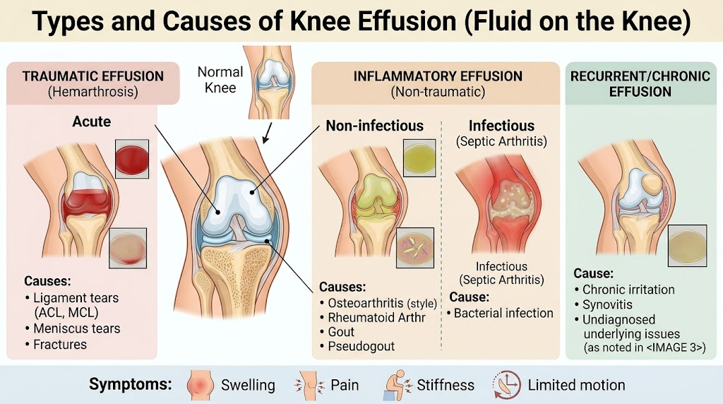
Causes of Knee Effusion by Type
The clinical etiology corresponds directly to the physical and biochemical properties of the aspirate:
- Serous Effusion: Most commonly secondary to knee osteoarthritis (especially prevalent in Egypt among individuals with elevated BMI and women over 50), non-specific synovitis, and rheumatoid arthritis. It develops gradually over days; patients notice swelling that worsens with prolonged standing or activity and subsides with rest. It tends to fluctuate alongside osteoarthritic flare-ups and is primarily managed conservatively.
- Hemorrhagic Effusion (Hemarthrosis): Develops rapidly within minutes to a few hours following trauma—acute post-traumatic swelling signifies a ligament rupture (e.g., ACL) or osteochondral fracture until proven otherwise. In young adults and athletes, football injuries represent the leading cause.
- Purulent Effusion: Develops over 24 to 48 hours, presenting with an intensely warm, erythematous, swollen knee accompanied by systemic fever and an inability to tolerate passive joint movement. It is typically caused by Staphylococcus aureus or other pathogens introduced via hematogenous spread or non-sterile intra-articular injections. Diabetic and immunocompromised patients face elevated susceptibility.
- Crystalline Effusion: Characterized by acute, abrupt attacks with severe pain, swelling, and localized erythema resembling infection, often with a normal systemic temperature. Gout is strongly linked to dietary habits rich in red meats and organ meats (common dietary patterns).
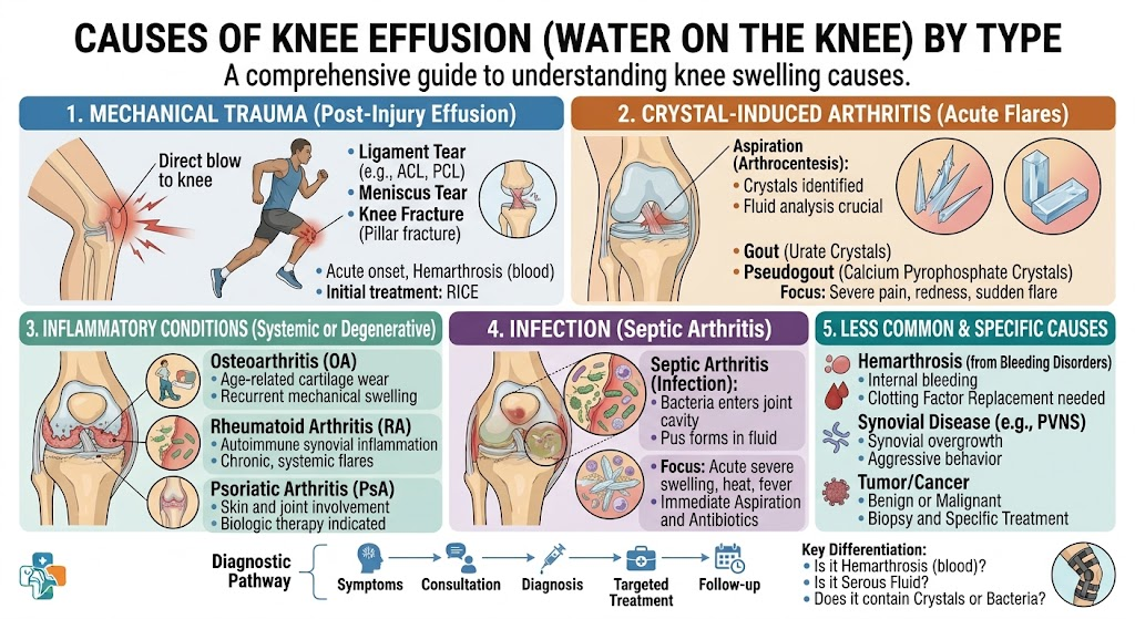
The Most Critical Types of Knee Effusion
Purulent effusion (septic arthritis) is unequivocally the most dangerous form. Bacterial enzymes and collagenases degrade articular cartilage within 24 to 48 hours—every hour of therapeutic delay inflicts irreversible chondral damage. Without immediate surgical lavage and intravenous antimicrobial coverage, patients face catastrophic joint destruction requiring reconstructive surgery or arthrodesis.
Differentiating Septic Arthritis from Acute Gout:
Both conditions present as a red, hot, swollen joint. However, septic arthritis typically involves systemic temperatures above 38.5°C, chills, and constitutional malaise, whereas gout presents primarily with localized joint agony and often normal body temperature. Definitive differentiation relies on synovial fluid analysis:
- A white blood cell (WBC) count exceeding 50,000/µL with polymorphonuclear dominance (>75–90%) and visible bacteria on Gram stain confirms septic arthritis.
- Negatively birefringent needle-shaped crystals under polarized light confirm gout.
- Whenever uncertainty exists, the joint is managed as an infection pending definitive microbiological culture results.
Hemorrhagic effusion ranks as the second most critical presentation. While blood does not degrade cartilage as rapidly as bacterial enzymes, it indicates structural mechanical disruption (ligamentous tears or intra-articular fractures) requiring prompt evaluation. Furthermore, intra-articular blood induces chemical synovitis and chondrocyte apoptosis if left unaddressed. Prompt arthrocentesis relieves elevated capsular pressure and allows macroscopic inspection (e.g., visible fat globules or lipohemarthrosis, which indicate an occult intra-articular fracture).
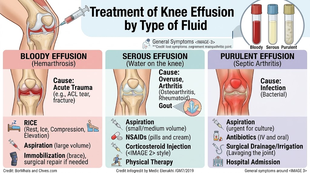
Treatment of Knee Effusion by Type
- Serous Effusion (Secondary to Osteoarthritis): Weight reduction (critical for decreasing joint load), targeted quadriceps strengthening, minimizing deep squatting and ground-level sitting, oral NSAIDs during flare-ups, and intra-articular hyaluronic acid or platelet-rich plasma (PRP) injections for recurrent symptomatic cases.
- Hemorrhagic Effusion: In-clinic joint aspiration to alleviate tension and inspect the aspirate, followed by knee MRI to assess ligamentous and meniscal integrity, proceeding to arthroscopic ligament reconstruction or meniscal repair/debridement when indicated.
- Purulent Effusion (Septic Arthritis): Zero delay permitted—immediate needle aspiration for cultures, followed by urgent arthroscopic or open joint irrigation and debridement on the same day, accompanied by intravenous antibiotic therapy initiated in-hospital (2–4 days) and transitioned to directed oral therapy to complete a 4–6 week course.
- Crystalline Effusion (Gout): Acute attack control using colchicine, NSAIDs, or intra-articular corticosteroids (3–7 days), followed by long-term serum urate-lowering therapy (such as allopurinol or febuxostat), along with dietary modifications: restricting red meats, organ meats, and high-fructose beverages, alongside optimized hydration.
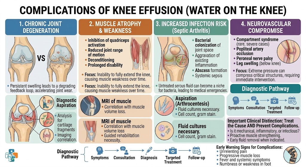
Complications of Knee Effusion
Complications vary based on the duration, volume, and underlying pathology of the effusion:
- Progressive Chondral Degradation: Untreated chronic effusions subject cartilage to continuous hydrostatic pressure and expose it to destructive inflammatory cytokines and matrix metalloproteinases, accelerating osteoarthritic progression even from seemingly benign serous fluid.
- Quadriceps Arthrogenic Muscle Inhibition and Atrophy: Pain and capsular distension inhibit neural drive to the quadriceps, leading to a 15–20% reduction in muscle mass within just two weeks of disuse, demanding prolonged rehabilitation to regain lost strength.
- Popliteal (Baker’s) Cyst: Excessive synovial fluid escapes through a one-way valve effect into the gastrocnemio-semimembranosus bursa, producing a posterior mass. Rupture of a Baker’s cyst discharges fluid into the calf, mimicking deep vein thrombosis (pseudothrombophlebitis).
- Catastrophic Joint Destruction (Purulent Only): Uncontrolled bacterial proliferation digests the chondral surface and subchondral bone, culminating in severe joint deformity that necessitates salvage total knee arthroplasty or arthrodesis.
- Chronic Intractable Infection: Inadequate or delayed debridement allows biofilm formation and persistent bacterial dormancy, predisposing the joint to recurrent infections and repeated operative washouts.
Frequently Asked Questions: Types of Knee Effusion
An authoritative clinical breakdown of synovial fluid classifications, hemarthrosis vs. serous effusion, diagnostic thresholds, and targeted treatments.
What are the four primary clinical classifications of knee effusion?
Synovial fluid analysis categorizes knee effusion into four major groups based on appearance, leukocyte count, and etiology:
• Non-Inflammatory: Clear, straw-colored transudate characteristic of osteoarthritis and mechanical overuse.
• Inflammatory: Cloudy, yellowish exudate driven by autoimmune arthritis or crystalline deposition.
• Septic: Opaque, purulent fluid indicative of acute intra-articular bacterial infection.
• Hemorrhagic (Hemarthrosis): Bloody fluid resulting from acute ligamentous trauma, capsular tears, or fractures.
What characterizes non-inflammatory knee effusion?
Non-inflammatory effusions are transparent, pale-yellow liquids with high stringency/viscosity (normal hyaluronic acid concentration) and a low white blood cell count (< 2,000 WBCs/μL with < 25% neutrophils). They are caused by mechanical wear rather than acute cellular immunity. Common conditions include primary knee osteoarthritis, chronic degenerative meniscal tears, osteochondritis dissecans, and joint overload.
What causes an inflammatory knee effusion, and how is it identified?
Inflammatory effusions appear translucent to turbid yellow with decreased viscosity and an elevated leukocyte count (2,000 to 50,000 WBCs/μL with > 50% polymorphonuclear neutrophils). Driven by active synovitis, they are associated with:
• Autoimmune Spondyloarthropathies: Rheumatoid arthritis, psoriatic arthritis, and reactive arthritis.
• Systemic Inflammatory Conditions: Systemic lupus erythematosus (SLE) and Lyme arthritis.
Patients typically report intense morning stiffness lasting longer than 45 to 60 minutes that improves with movement.
What is septic knee effusion, and why is it considered a medical emergency?
Septic effusion represents an acute bacterial joint infection (most commonly Staphylococcus aureus or Streptococcus species). The fluid is opaque, dense, or frankly purulent with > 50,000 to 100,000+ WBCs/μL (> 75–90% neutrophils), low glucose, and high lactate. It is a critical surgical emergency because bacterial collagenases, metalloproteinases, and lysosomal enzymes rapidly destroy articular cartilage within 24 to 48 hours, requiring urgent surgical drainage and IV antibiotics.
What is hemarthrosis, and what are its most common traumatic causes?
Hemarthrosis is the intra-articular accumulation of frank blood, which triggers rapid, tense joint swelling within 1 to 4 hours post-trauma. The primary etiologies include:
• Anterior Cruciate Ligament (ACL) Tears: Responsible for over 70% of acute traumatic hemarthroses.
• Patellar Dislocation / Subluxation: Involves tearing of the medial patellofemoral ligament (MPFL).
• Peripheral Meniscal Tears: Tears in the vascularized ‘red-red’ outer third of the meniscus.
• Coagulopathies: Non-traumatic bleeding in hemophilia or patients on anticoagulant therapy.
What is lipohemarthrosis, and what does its presence indicate?
Lipohemarthrosis occurs when bone marrow fat globules mix with blood inside the joint capsule. Because fat is less dense than blood, it floats, producing a characteristic fat-fluid level on cross-table horizontal-beam X-rays, CT, or MRI. Lipohemarthrosis is pathognomonic for an intra-articular fracture (such as a tibial plateau fracture, distal femoral fracture, or patellar fracture) that has breached the cortical bone and medullary cavity.
How are crystal-induced knee effusions (gout and pseudogout) differentiated?
Both present as sudden, exquisitely painful inflammatory effusions with marked cutaneous warmth, but they are definitively differentiated under compensated polarized light microscopy:
• Gout: Demonstrates needle-shaped monosodium urate (MSU) crystals that show strong negative birefringence (turning yellow when aligned parallel to the compensator axis).
• Pseudogout (CPPD): Demonstrates pleomorphic, rhomboid calcium pyrophosphate dihydrate crystals showing weak positive birefringence (turning blue when aligned parallel), often accompanied by radiographic chondrocalcinosis.
What is pigmented villonodular synovitis (PVNS), and what type of effusion does it cause?
Pigmented Villonodular Synovitis (PVNS), classified as a tenosynovial giant cell tumor (TGCT), is a benign proliferative disease of the synovial membrane. It characteristically produces recurrent, spontaneous serosanguinous or dark brownish-red effusions in young adults without prior trauma. The dark discoloration stems from extensive hemosiderin deposition within the hyperplastic synovial tissue, which produces characteristic blooming artifacts on gradient-echo MRI sequences.
What is a chylous knee effusion, and how rare is it?
Chylous knee effusion (chylarthrosis) is an exceptionally rare presentation where joint fluid appears milky white or opalescent. Laboratory testing confirms markedly elevated triglycerides and cholesterol concentrations. It results from traumatic rupture or congenital malformation of periarticular lymphatic vessels, severe lymphatic obstruction, or systemic lipid metabolism disorders, requiring lymphatic imaging and non-weight-bearing management.
How does identifying the exact type of knee effusion guide definitive management?
Accurate fluid classification directs targeted clinical intervention:
• Non-inflammatory: Physical therapy, quadriceps strengthening, weight loss, and viscosupplementation or PRP.
• Inflammatory: Systemic rheumatologic disease-modifying therapies (DMARDs) or colchicine/NSAIDs for crystal flares.
• Septic: Immediate emergent arthroscopic arthrotomy/lavage and culture-directed parenteral antibiotic regimens.
• Traumatic Hemarthrosis: Diagnostic MRI followed by arthroscopic ligament reconstruction or meniscal repair.