Knee effusion is one of the most common reasons for visiting an orthopedic clinic in Egypt. It is not a disease in itself, but rather a clinical sign indicating an intra-articular pathology that requires accurate diagnosis. The accumulated fluid may be excess synovial fluid (as seen in osteoarthritis and inflammatory conditions), blood (in trauma and fractures), or pus (in septic arthritis, which is a medical emergency). Identifying the nature of the fluid and its underlying etiology is the first step toward effective management. This article outlines the various causes of knee effusion, when arthrocentesis (fluid aspiration) is indicated, and when conservative management is sufficient.
What is Knee Effusion?
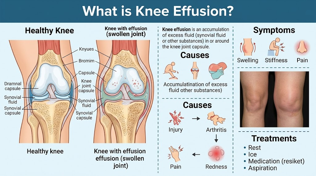
Knee effusion refers to the abnormal accumulation of excess fluid within the knee joint capsule. A normal knee joint contains approximately 2 to 3 mL of synovial fluid, which lubricates the articulating surfaces and nourishes the avascular articular cartilage. Following inflammation or injury, the inner synovial lining becomes hyperactive and produces excessive effusion within the joint cavity, resulting in swelling, pain, and restricted mobility.
The volume of effusion can range from 10–20 mL (mild, presenting as subtle swelling) to 80–100 mL or more (severe, presenting as a tense, balloon-like swelling around the joint). Massive effusion prevents full knee flexion because the fluid occupies internal intra-articular space and elevates capsular pressure. Clinical examination readily detects effusion through tests such as the patellar tap test (ballottement), where pressing the patella downward produces a floating or tapping sensation against the underlying femoral condyle, and the fluid wave (bulge) test, where displacing fluid from one compartment reveals a visible fluid wave on the contralateral side.
Causes of Knee Effusion
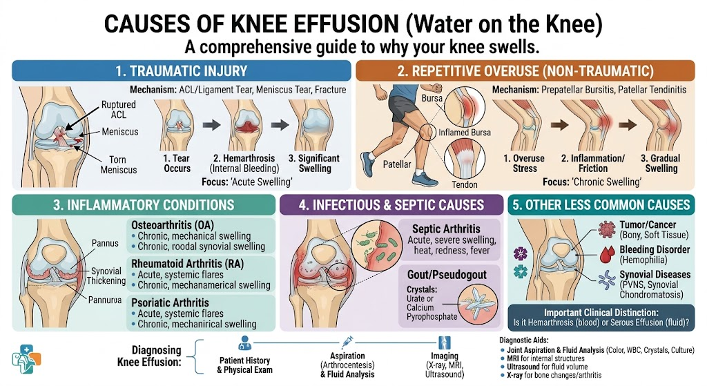
The underlying causes are clinically categorized by synovial fluid gross appearance and laboratory characteristics:
Serous / Translucent Yellow: Knee osteoarthritis (the most common cause in Egypt), non-infectious synovitis, and rheumatoid arthritis.
Urgent evaluation required to identify structural trauma
Purulent (Turbid)
Opaque, yellowish-green, malodorous
Bacterial septic arthritis
Emergency; urgent joint washout/debridement within hours
Crystalline
Cloudy yellow; microcrystals present
Gout, calcium pyrophosphate deposition (CPPD)
Sub-acute/urgent; requires prompt medical treatment
Symptoms of Knee Effusion
Clinical manifestations vary according to the volume and primary cause of the effusion:
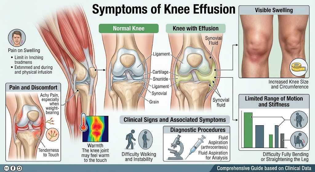
Mild Effusion: Subtle peripatellar swelling, a sensation of intra-articular fullness, and morning stiffness.
Moderate Effusion: Obvious joint swelling restricting full terminal flexion, accompanied by pain during weight-bearing and stair climbing.
Severe Effusion: Tense, visibly distended joint with marked mechanical limitation and inability to bear weight.
Septic Arthritis: Marked swelling accompanied by localized erythema, increased skin temperature, fever, and severe pain during minimal passive motion.
Key Signs & Symptoms:
Peripatellar Swelling: The earliest visible sign; loss of normal bony landmarks around the patella and obliteration of the parapatellar gutters.
Restricted Range of Motion: Terminal flexion becomes painful or mechanically blocked as internal fluid volume distends the capsule.
Activity-Related Pain: Prolonged walking, navigating stairs, and deep squatting exacerbate both pain and intra-articular pressure.
Subjective Instability: Sensation of the knee “giving way,” particularly when an underlying ligamentous disruption is present.
Local Warmth and Erythema: Critical red flags suggesting septic arthritis or acute crystal arthropathy, warranting emergency evaluation.
Can One Live with Knee Effusion?
Tolerating knee effusion depends entirely on its severity and underlying cause. Mild, recurrent effusion secondary to chronic osteoarthritis can often be managed conservatively through weight reduction, quadriceps strengthening, and avoiding excessive mechanical load (such as repeated stair climbing and deep kneeling). In contrast, persistent large-volume effusions require definitive diagnosis, as chronic inflammatory fluid contains catabolic enzymes that degrade articular cartilage. Any effusion presenting with localized warmth and erythema indicates potential infection and cannot be left unaddressed.
For patients in Egypt managing osteoarthritic effusion, recurrences can be substantially reduced by:
Weight Reduction: Losing 5–10 kg decreases mechanical knee load by 15–50 kg per step.
Targeted Quadriceps Strengthening: Daily low-impact exercises enhance dynamic joint stability and unload the cartilage.
Modifying Daily Postures: Minimizing deep squatting and floor sitting.
Cryotherapy: Applying ice packs for 15 minutes after demanding physical tasks.
If an effusion recurs more than 3 to 4 times annually or produces significant distension requiring repeated drainage, finding and addressing the primary joint pathology is essential rather than relying solely on recurrent aspirations.
Treatment of Knee Effusion
Management involves symptom-directed relief and targeted treatment of the underlying condition.
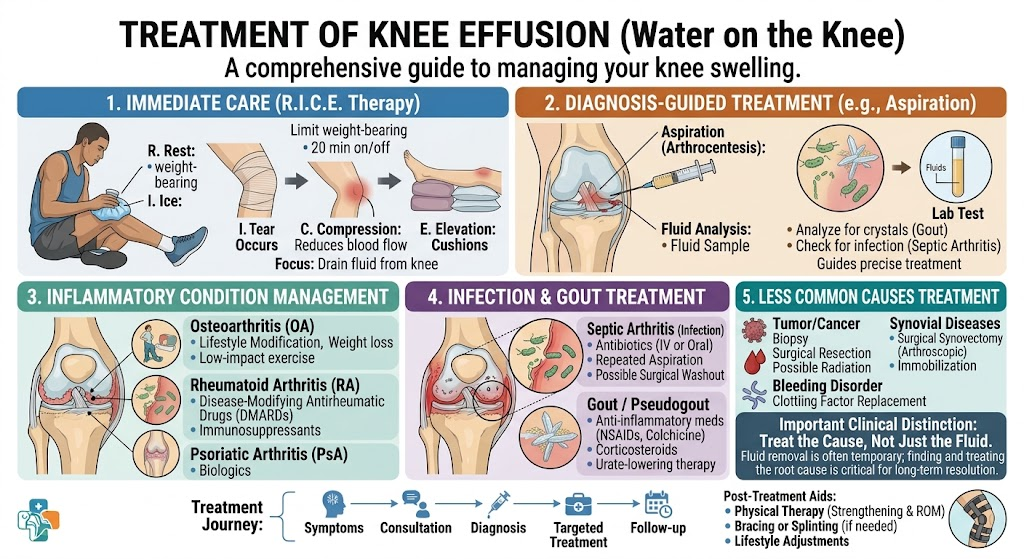
Symptomatic Management:
Rest, ice packs (15–20 minutes every few hours), elevation, and light compressive bandaging.
Non-steroidal anti-inflammatory drugs (NSAIDs such as ibuprofen or diclofenac).
Arthrocentesis (needle drainage) for tense, painful effusions, with fluid sent for laboratory analysis (cell count, Gram stain, culture, and polarized light microscopy).
Etiology-Specific Treatment:
Osteoarthritis: Weight optimization, physical therapy, NSAIDs, and intra-articular hyaluronic acid or platelet-rich plasma (PRP) injections. Refractory cases may require arthroscopic intervention or total knee arthroplasty depending on radiographic staging.
Ligamentous or Meniscal Tears: Initial drainage of hemarthrosis followed by MRI evaluation, followed by arthroscopic repair or reconstruction as indicated.
Gout / Pseudogout: Urate-lowering therapy, colchicine or short-course corticosteroids for acute flares, and dietary adjustments (moderating purine-rich foods and high-fructose beverages).
Rheumatoid Arthritis: Disease-modifying antirheumatic drugs (DMARDs) and biologic therapy under rheumatologic guidance, with judicious intra-articular corticosteroid injections during flare-ups.
Septic Arthritis (Orthopedic Emergency): Immediate joint aspiration for synovial analysis and cultures, urgent arthroscopic or open joint debridement/lavage, and 4–6 weeks of targeted intravenous and oral antimicrobial therapy. Delayed intervention risks irreversible chondral destruction.
Key Clinical Consideration: Aspiration alone without treating the root cause typically leads to fluid re-accumulation within days or weeks. Routine, unguided fluid drainage without identifying the driver accelerates joint compromise. Synovial fluid analysis at the initial aspiration dictates the diagnostic pathway: inflammatory fluid warrants a rheumatologic workup, hemarthrosis requires advanced imaging (MRI) for structural injury, and purulent fluid necessitates emergency surgical debridement.
Clinical Reference Guide
Frequently Asked Questions: Knee Effusion
An authoritative clinical overview of fluid accumulation causes, physical examination tests, synovial fluid analysis, infectious red flags, and treatments.
What is knee effusion, and how does it differ from periarticular edema?
Knee effusion—colloquially referred to as “water on the knee”—is the abnormal buildup of excess fluid strictly within the synovial capsule of the joint. In contrast, periarticular edema represents diffuse interstitial fluid retention in the skin and subcutaneous tissues outside the capsule. Knee effusion typically creates a tense sensation of fullness in the suprapatellar pouch, a ballotable patella, and mechanical stiffness that limits terminal flexion.
What does the timing of knee swelling indicate after an acute injury?
The temporal onset of swelling provides vital diagnostic clues:
• Immediate Swelling (< 1–4 hours): Points to acute hemarthrosis (gross blood within the joint), most frequently caused by an ACL tear (over 70% of cases), patellar dislocation, or osteochondral fracture.
• Delayed Swelling (12–24+ hours): Represents serous synovial fluid produced in response to mechanical irritation, typical of meniscal tears, mild ligamentous sprains, or early osteoarthritis flares.
What are the primary clinical tests used to detect a knee effusion?
Clinicians assess knee effusion through physical examination maneuvers:
• Bulge / Sweep Test (Stroke Test): Highly sensitive for minimal fluid (under 4–8 mL). The clinician sweeps fluid upward from the medial joint line into the suprapatellar pouch, then strokes the lateral side downward; a visible fluid wave returning to the medial sulcus indicates a positive test.
• Patellar Tap (Ballottement) Test: Used for moderate-to-large effusions. The examiner compresses the suprapatellar pouch to gather fluid beneath the patella and pushes the patella downward; a palpable tap or floating sensation against the femoral condyles confirms significant fluid accumulation.
When is knee joint aspiration (arthrocentesis) necessary?
Arthrocentesis serves both diagnostic and therapeutic purposes:
• Diagnostic Utility: Critical when the cause of acute swelling is uncertain, especially to distinguish between infection, crystal arthropathy (gout/pseudogout), and hemarthrosis with fat droplets (lipohemarthrosis indicative of an occult fracture).
• Therapeutic Relief: Evacuating tense, high-pressure effusion provides instant relief from pain and decompresses the capsule, facilitating clinical range-of-motion testing and physical rehabilitation.
How does synovial fluid analysis distinguish between different knee pathologies?
Aspirated fluid is analyzed for clarity, color, cell counts, and crystal microscopy:
• Normal / Non-Inflammatory (e.g., Osteoarthritis): Clear, pale yellow, WBC < 2,000/μL.
• Inflammatory (e.g., Rheumatoid, Gout, Pseudogout): Translucent to cloudy yellow, WBC 2,000–50,000/μL with specific crystals visible under polarized light.
• Septic (Bacterial Infection): Turbid, opaque, or purulent, WBC > 50,000/μL (> 75–90% neutrophils) with positive Gram stain and culture.
• Hemorrhagic: Frank red blood, indicative of ligament ruptures, vascular capsular tears, or intra-articular fractures.
What red flag symptoms indicate an infected knee (septic arthritis)?
Septic arthritis is an orthopedic emergency. Warning signs include:
• High fever, rigors, or systemic malaise.
• Rapid joint distension with marked cutaneous erythema and warmth.
• Intense, severe pain provoked even by gentle passive motion of the joint.
• Absolute inability to bear weight.
Immediate emergency department evaluation and synovial aspiration are required to prevent permanent enzymatic destruction of articular cartilage.
Why is a Baker’s cyst (popliteal cyst) associated with knee effusion?
A Baker’s cyst is a secondary phenomenon rather than a primary disease. There is an anatomical communication between the knee joint capsule and the gastrocnemius-semimembranosus bursa in the popliteal fossa. When an intra-articular condition (such as a degenerative meniscus tear or osteoarthritis) generates excess synovial effusion, elevated hydrostatic pressure pushes fluid through a one-way valve mechanism into the bursa, causing posterior fullness and tightness during knee extension.
What is the initial conservative management protocol for knee effusion?
Non-infectious, mechanical, or inflammatory effusions are managed via conservative measures:
• Rest: Avoiding provocative axial impact and prolonged standing.
• Ice: Applying cryotherapy for 15–20 minutes every 2 to 3 hours to induce local vasoconstriction.
• Compression: Using an elastic bandage or compression sleeve to minimize intra-capsular fluid extravasation.
• Elevation: Propping the leg above heart level to assist lymphatic drainage.
• Pharmacotherapy: Short-term oral or topical NSAIDs to downregulate synovial prostaglandin production.
How does chronic knee effusion affect surrounding muscles?
Joint effusion induces a reflex neurophysiological shutdown known as Arthrogenic Muscle Inhibition (AMI). As little as 20 to 30 mL of fluid within the knee capsule stimulates intra-articular mechanoreceptors, sending continuous inhibitory feedback through spinal pathways that prevents the central nervous system from fully recruiting the quadriceps muscles (especially the vastus medialis oblique). This leads to rapid disuse atrophy, functional weakness, and joint instability if the effusion is not addressed.
When does a knee effusion require arthroscopic surgery?
Knee arthroscopy is indicated when persistent or recurrent effusion is driven by structural internal derangements that cannot heal with conservative therapy:
1. Symptomatic, unstable meniscal tears (e.g., bucket-handle or radial tears causing mechanical catching).
2. Complete cruciate ligament tears (ACL/PCL) creating recurrent rotational instability and secondary effusions.
3. Intra-articular loose bodies (detached bone or cartilage fragments causing locking).
4. Chronic hypertrophic synovitis refractory to medical therapy (requiring arthroscopic synovectomy).