
Knee pain is one of the most common medical complaints in Egypt, and many cases respond well to treatment of knee pain in home without the immediate need for clinical intervention. In this comprehensive medical guide, you will learn about the primary causes of knee pain and how to differentiate between sudden pain requiring urgent evaluation and pain manageable at home. You will discover what clinical evidence says regarding walking and knee pain, the fastest evidence-based home protocols, how to overcome chronic knee pain permanently through a multimodal regimen of targeted exercises, over-the-counter pharmacology, and lifestyle modifications, as well as the critical red flags necessitating an immediate specialist consultation.
Most Common Causes of Knee Pain
Knee pain is not a single diagnosis—it comprises a heterogeneous spectrum of conditions, each possessing distinct pathophysiological etiologies and therapeutic pathways. Establishing an accurate anatomical diagnosis represents the cornerstone of effective management.
- Knee Osteoarthritis (OA): The most common etiology in individuals over 50 years of age, characterized by progressive articular cartilage degeneration, joint space narrowing, and chronic aching pain exacerbated by cold exposure, weight-bearing, and prolonged ambulation.
- Meniscal Tears: Frequently seen in young athletes following sudden rotational torsional forces; presents with acute localized joint-line tenderness, effusion, and mechanical sensations such as catching or true joint locking.
- Anterior Cruciate Ligament (ACL) Ruptures: Typically occur secondary to rapid deceleration, cutting maneuvers, or improper landing; characterized by immediate acute pain, a perceptible audible “pop,” rapid hemarthrosis, and subjective knee instability or “giving way.”
- Patellofemoral Pain Syndrome (PFPS / “Runner’s Knee”): Prevalent among adolescents, young adults, and female athletes; manifests as peripatellar or retropatellar anterior knee pain aggravated by stair descent/ascent, prolonged squatting, and prolonged sitting with knees flexed (“theater sign”).
- Prepatellar or Pes Anserine Bursitis: Produces focal periarticular tenderness, localized swelling, and prominent nocturnal discomfort with palpable local warmth.
- Patellar Tendinopathy (Jumper’s Knee): Results from repetitive mechanical overload and microtrauma to the patellar tendon insertion, common in jumping and running sports.
- Post-Traumatic Skeletal Disruptions: Articular fractures (e.g., tibial plateau, distal femur, patella) and acute joint dislocations require immediate emergency orthopaedic intervention.

Sudden Knee Pain: When Is Urgent Action Required?
Acute, sudden knee pain differs fundamentally in pathophysiology and management from gradual, progressive chronic discomfort.
- Sudden Pain Following Acute Mechanical Trauma or Twisting: Discontinue all weight-bearing immediately and apply the PRICE protocol.
- Sudden Pain Without Clear Trauma Accompanied by Rapid Swelling (within hours): Strongly suggests intra-articular hemarthrosis, high-grade soft-tissue disruption, or acute septic arthritis, mandating prompt orthopaedic evaluation.
- Sudden Pain with an Audible “Pop” and Inability to Bear Weight: Highly indicative of an acute ACL or collateral ligament disruption; attend an orthopaedic clinic or emergency department the same day.
- Sudden Pain Accompanied by Severe Local Erythema, Warmth, and Systemic Fever: Strongly suspicious for acute septic arthritis—a genuine orthopaedic emergency requiring urgent joint aspiration and intravenous antimicrobial therapy.
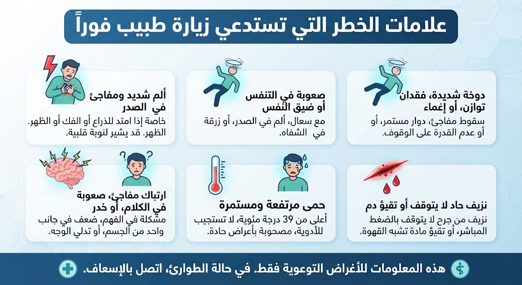
Red Flags Requiring an Immediate Medical Consultation
The emergence of any of the following clinical signs indicates that home care is inadequate, and continued reliance on self-treatment risks structural joint compromise or delayed intervention.
| Warning Sign (Red Flag) | Suspected Pathology | Required Clinical Action |
| Complete inability to bear weight on the extremity | Acute fracture or full-thickness ligamentous disruption | Same-day urgent evaluation / Emergency room |
| Rapid joint effusion developing within 1–2 hours | Acute hemarthrosis / Complete ACL rupture | Urgent orthopaedic consultation within 24 hours |
| Marked effusion with localized erythema, heat, and high fever | Acute septic arthritis | Immediate emergency evaluation within hours |
| True joint locking (inability to achieve full extension) | Displaced bucket-handle meniscal tear / Loose body | Urgent surgical orthopaedic evaluation |
| Gross visible anatomical deformity of the joint | Knee dislocation or displaced intra-articular fracture | Immediate emergency department reduction/care |
| Intractable, severe nocturnal pain unrelieved by resting | Neoplastic lesion or active systemic inflammatory arthropathy | Comprehensive clinical evaluation within 1 week |
| Persistent localized pain lasting > 3 weeks despite conservative care | Chronic occult meniscal/chondral tear or advanced OA | Routine specialist orthopaedic outpatient appointment |
Does Walking Aggravate Knee Pain?
The established scientific consensus is definitive: regular, low-impact, moderate walking is profoundly beneficial for the vast majority of knee conditions—most notably knee osteoarthritis—and is not harmful. The Osteoarthritis Research Society International (OARSI) 2019 guidelines (reaffirmed in recent updates) categorize structured land-based walking as a core, comprehensive treatment modality for knee OA, irrespective of baseline disease severity.
- Clinical Trial Data: In a cohort of over 1,200 individuals with radiographically confirmed knee osteoarthritis from the Osteoarthritis Initiative (OAI), regular walkers demonstrated a statistically significant reduction in the development of recurrent frequent knee pain compared to non-walkers.
- Delaying Arthroplasty: Data published through OARSI indicate that increasing daily structured walking by just 5 minutes is associated with a 16% reduction in the risk of undergoing total knee arthroplasty over long-term follow-up. Walking enhances cyclic hydrostatic pressure within the joint, facilitating synovial fluid circulation and chondrocyte nutrition, strengthening stabilizing periarticular musculature, and downregulating chronic intra-articular pro-inflammatory cytokines.
- When Is Walking Contraindicated? In the acute inflammatory phase accompanied by tense effusion or immediately following acute mechanical trauma. In such scenarios, initiate relative joint protection for the first 24–72 hours prior to reintroducing gradual protected ambulation. In severe, end-stage (Kellgren-Lawrence Grade IV) osteoarthritis, walking remains beneficial within physiological tolerance limits, structured in shorter bouts and supported by an assistive device (e.g., contralateral walking cane) to unload joint reaction forces.
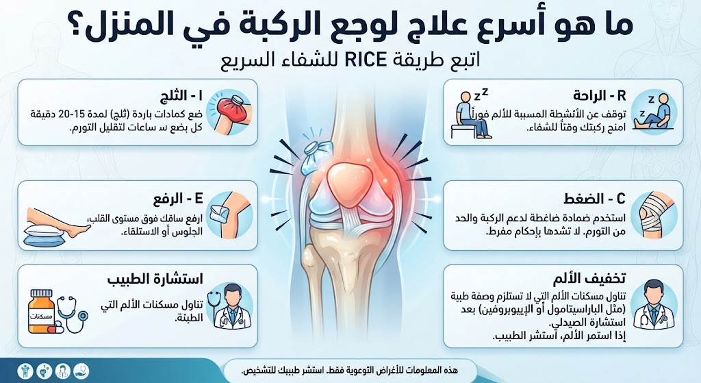
What Is the Fastest treatment of knee pain in home?
The PRICE protocol remains the most rapid, evidence-supported first-line strategy to manage acute knee pain at home, yielding symptomatic improvement within 24 to 48 hours when applied correctly.
- Protection: Shield the joint from additional mechanical trauma using external support (e.g., hinged or soft neoprene knee sleeve) and avoid provocative movements without resorting to absolute immobilization. Controlled gentle motion preserves local peripheral vascular flow.
- Rest (Relative Rest): Modify daily physical activity rather than committing to strict bed rest. Low-load light ambulation is acceptable, while running, jumping, heavy lifting, and deep squatting are strictly avoided during acute flares.
- Ice (Cryotherapy): Apply cold packs wrapped in a thin protective cloth for 15–20 minutes every 2–3 hours during the initial 48–72 hours. Cryotherapy induces local vasoconstriction, limits secondary metabolic cellular injury, and rapidly dampens local edema, inflammation, and nociceptive nerve conduction.
- Compression: Utilize a snug, breathable elastic bandage or knee compression sleeve to limit interstitial fluid extravasation, support joint proprioception, and contain acute capsular swelling.
- Elevation: Prop the affected limb above the level of the heart whenever seated or recumbent to promote gravity-assisted lymphatic drainage and venous return.
- Thermotherapy: Heat applications (warm compresses, hydrocollator packs) are strictly indicated after 48–72 hours, once the acute inflammatory phase has subsided. Superficial heat increases local tissue perfusion, alleviates secondary myofascial guarding, and restores soft-tissue compliance. Applying heat during acute inflammatory swelling is contraindicated, as it induces vasodilation and exacerbates edema.
- Oral Anti-inflammatory Pharmacotherapy: Short courses (5–7 days) of oral NSAIDs—such as diclofenac (50 mg twice daily with food) or celecoxib (200 mg once daily)—provide rapid reduction in inflammatory prostaglandins and joint effusion in appropriately screened patients without gastrointestinal, renal, or cardiovascular contraindications.
- Topical NSAIDs: Topical formulations such as Diclofenac Diethylamine Gel (1–2%) applied directly over the knee 2–4 times daily offer local anti-inflammatory efficacy comparable to oral agents with substantially fewer systemic and gastrointestinal side effects.
Home Exercises for Knee Pain Relief
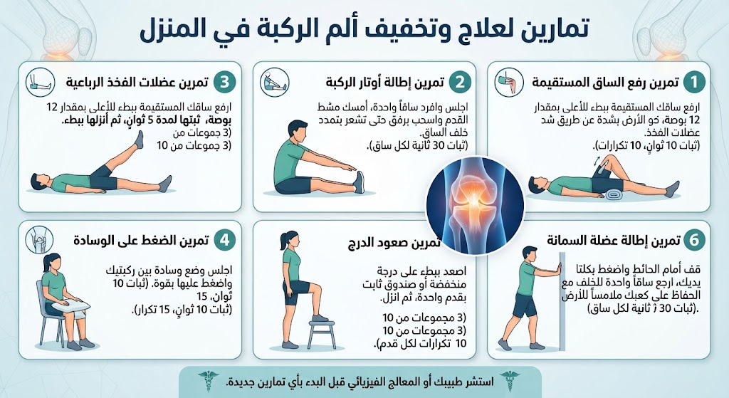
Progressive resistance training focused on the quadriceps and hip abductor/gluteal musculature delivers the highest long-term structural and functional efficacy. Robust clinical literature demonstrates substantial reductions in pain scores and functional gains within 6 to 12 weeks of consistent execution.
                  HOME REHABILITATION PROGRESSION
┌───────────────────────┐     ┌───────────────────────┐     ┌───────────────────────┐
│ Straight Leg Raise    │ ──> │ Wall Sit & Glute      │ ──> │ Hamstring / ITB       │
│ (Isolates Quad        │     │ Bridge (Kinetic Chain │     │ Stretching (Flexibility│
│ without Joint Stress) │     │ & Peripelvic Stability│     │ & Joint Decompression)│
└───────────────────────┘     └───────────────────────┘     └───────────────────────┘
- Straight Leg Raise (SLR):
- Execution: Lie supine on a firm surface. Flex the unaffected limb so the foot rests flat on the floor. Keeping the affected knee locked in full extension, elevate the limb smoothly to the level of the contralateral flexed knee. Hold isometrically for 3 seconds, then lower under control.
- Prescription: 3 sets of 15 repetitions daily.
- Mechanism: Strengthens the rectus femoris and vastus medialis without introducing patellofemoral compressive contact stresses.
- Heel Slides:
- Execution: Lie supine with both legs extended. Slowly slide the heel of the affected limb toward the buttocks, flexing the knee within a comfortable, pain-free range of motion, then smoothly slide back to full extension.
- Prescription: 3 sets of 15 repetitions daily.
- Mechanism: Safely restores active-assisted flexion/extension range of motion in stiff or post-inflammatory joints.
- Wall Sit:
- Execution: Stand with your back resting flat against a smooth wall, feet shoulder-width apart and roughly two feet forward. Slowly slide downward until knees achieve an angle of approximately 45° (do not exceed 90° of flexion). Hold the static position for 10–30 seconds, then return to standing.
- Prescription: 3–4 sustained isometric holds daily.
- Mechanism: Provides closed-kinetic-chain isometric quadriceps strengthening, stabilizing the extensor mechanism.
- Glute Bridge:
- Execution: Lie supine with both knees flexed at 90° and feet flat on the ground. Contract the core and gluteal musculature, lifting the pelvis until the thighs, hips, and torso form a straight diagonal line. Hold for 5 seconds at the top, then descend slowly.
- Prescription: 3 sets of 15 repetitions daily.
- Mechanism: Strengthens the gluteus medius and maximus. Adequate peripelvic strength controls femoral internal rotation and adduction, offloading dynamic knee valgus forces.
- Standing Hamstring Stretch:
- Execution: Stand upright, placing the heel of the affected limb forward on an elevated platform or low stool with the knee straight. Hinge forward at the hips while maintaining a neutral spine until a gentle, tolerable stretch is felt along the posterior thigh. Maintain static stretch for 30 seconds.
- Prescription: 3 repetitions per limb daily.
- Mechanism: Resolves hamstring hypertonicity, reducing retropatellar joint reaction forces during weight-bearing stance.
Eliminating Knee Pain Permanently
Permanent resolution of chronic knee pain demands an integrated, multimodal therapeutic approach rather than reliance on short-term analgesia. Analgesics provide temporary symptomatic relief, whereas targeted mechanical offloading, kinetic chain re-education, and metabolic optimization target the underlying pathology.
- Biomechanical Weight Management: Weight optimization is the most potent disease-modifying intervention in lower-extremity osteoarthritis. Biomechanical studies confirm that every single kilogram of excess body weight exerts 4 kilograms of cumulative joint reaction force across the tibiofemoral joint during ambulation. Achieving a modest 5% to 10% reduction in total body weight yields clinically meaningful reductions in joint pain, markers of systemic inflammation, and radiographic disease progression within months.
- Kinetic Chain Conditioning: Long-term adherence to quadriceps, hamstring, and hip musculature strengthening maintains dynamic joint stability, decreasing shearing forces on damaged articular cartilage.
- Ergonomic Footwear: Utilize motion-control or well-cushioned supportive footwear equipped with shock-absorbing soles, reducing peak vertical ground reaction forces transmitted to the knee by up to 25% during gait. Avoid sustained barefoot ambulation across unyielding, rigid surfaces (e.g., hard tile or concrete floors).
- Activity and Postural Modification: Minimize deep hyperflexion maneuvers, including deep squatting, prolonged floor sitting, and unnecessary stair ascents/descents. When navigating stairs, make systematic use of handrails to unload the joint, and modify home furniture to favor slightly elevated seating surfaces.
- Evidence-Based Nutraceuticals: Daily supplementation with crystalline Glucosamine Sulfate and Chondroitin Sulfate provides modest, high-safety symptomatic benefits for pain reduction and joint space preservation in approximately 30–40% of patients with mild-to-moderate osteoarthritis after 3 to 6 months of continuous use. Correction of documented hypovitaminosis D and systemic calcium deficiencies is mandatory to preserve subchondral bone integrity.
- Targeted Image-Guided Injections: Ultrasound-guided corticosteroid injections provide rapid relief in acute inflammatory flares; intra-articular high-molecular-weight hyaluronic acid viscosupplementation restores synovial lubrication and shock absorption in mild-to-moderate osteoarthrosis; autologous Platelet-Rich Plasma (PRP) offers biologically regenerative, anti-inflammatory support in refractory chondral injuries and tendinopathies.
- Indications for Surgical Intervention: When structured conservative protocols fail to achieve functional restoration after 6 months in cases of severe radiographic joint destruction (Kellgren-Lawrence Grade III/IV), or in the presence of acute high-grade mechanical pathology (irreducible bucket-handle meniscal tears, complete multi-ligamentous ruptures), orthopaedic surgical evaluation for arthroscopic reconstruction or Total Knee Arthroplasty (TKA) is warranted.
Clinical Treatment Pathways and Strategic Timelines
| Therapeutic Objective | Optimal Modality / Clinical Strategy | Expected Treatment Horizon |
| Acute Pain and Effusion Control | PRICE protocol + Short-course oral/topical NSAIDs | 3–7 days |
| Subacute Swelling & Edema Resolution | Progressive isometric exercise + Supervised weight loss | 4–8 weeks |
| Structural Cartilage Preservation | Low-impact walking + Kinetic chain closed-chain strengthening | Indefinite / Continuous |
| Mild-to-Moderate Osteoarthritis | Image-guided viscosupplementation / PRP + Physical therapy | 3–6 months |
| End-Stage / Refractory Joint Pathology | Orthopaedic evaluation for Total Knee Replacement (TKA) | After failing ≥ 6 months of conservative care |
Book an Appointment with Prof. Dr. Ibrahim Abdel Moneim
Prof. Dr. Ibrahim Abdel Moneim, Professor of Orthopedic and Joint Surgery at Ain Shams University and Visiting Professor of Orthopedic Surgery at Ohio State University, provides advanced differential clinical examinations and high-resolution ultrasound evaluations to formulate customized, comprehensive treatment protocols for complex knee and shoulder conditions.
Clinics are located in:
- Nasr City
- New Cairo (Fifth Settlement)
- Mohandessin
- El-Batal Ahmed Abdel Aziz Street
Frequently Asked Questions: Treatment of Knee Pain at Home
What are the immediate home steps to relieve sudden knee pain and swelling?
When should you use cold packs versus warm compresses for knee pain?
What simple home exercises help relieve knee pain safely?
Do topical analgesics and anti-inflammatory creams help treat knee pain at home?
How do excess weight and lifestyle factors impact knee pain, and how can they be managed at home?
Can knee sleeves or medical braces be worn continuously at home?
What are common mistakes people make when treating knee pain by themselves?
How can you modify your home environment to be more comfortable for knee pain?
When do home remedies become insufficient, requiring an immediate orthopedic consultation?
What medical evaluations does a doctor perform if knee pain does not improve with home treatment?
references
- OARSI 2024 — Walking and Exercise as Core Treatment for Knee OA
- Osteoarthritis Initiative Cohort — Walking for Exercise and Knee OA Progression
- GoodRx — Home Remedies for Knee Pain: Evidence-Based Guide
- Apollo 247 — Natural Treatments for Knee Pain (2026)
- EmergeOrtho — 10 Tips for Managing Knee Pain at Home (2026)
- Home-Based vs Center-Based Exercise for Knee Osteoarthritis: Systematic Review with Meta-Analysis — Frontiers in Public Health (2024)
- Supervised or Home-Based? Best Exercise Approach for Knee Osteoarthritis: Systematic Review & Meta-Analysis — J Clinical Medicine (2025)
- Efficacy of Home-Based Exercise for Pain and Disability in Knee Osteoarthritis: Systematic Review & Meta-Analysis — BMC Musculoskeletal Disorders (2024)
- Home-Based Exercise for Knee & Hip Osteoarthritis: Systematic Review — PMC (2024)
- Knee Pain Self-Care: RICE Method, Ice, Heat & Exercises — Sutter Health (2024)
- How to Treat Knee Pain at Home: Ice, Heat, Exercises & Medications — UPMC (2022)
- Managing Knee Pain at Home: RICE, NSAIDs, Physical Therapy — UPMC (2025)
- Home Care for Knee Pain: Ice, Heat, Compression & Elevation — Hawaii Pacific Health (2024)
- How to Reduce Knee Pain at Home: RICE, Ginger, Lifestyle Changes — eMediHealth
- Evaluation and Treatment of Knee Pain: A Review — JAMA (2023)