
Symptoms of collateral knee ligament tears and their three grades, causes, diagnosis, conservative treatment options, medications, injections, and surgery — Dr. Ibrahim Shaarawi, Cairo.
What are the collateral knee ligaments?
The collateral knee ligaments are two bands that stabilize the knee from its sides: the Medial Collateral Ligament (MCL) on the inner side, and the Lateral Collateral Ligament (LCL) on the outer side. They prevent the knee from shifting inward or outward and serve as the first line of defense against lateral injuries.
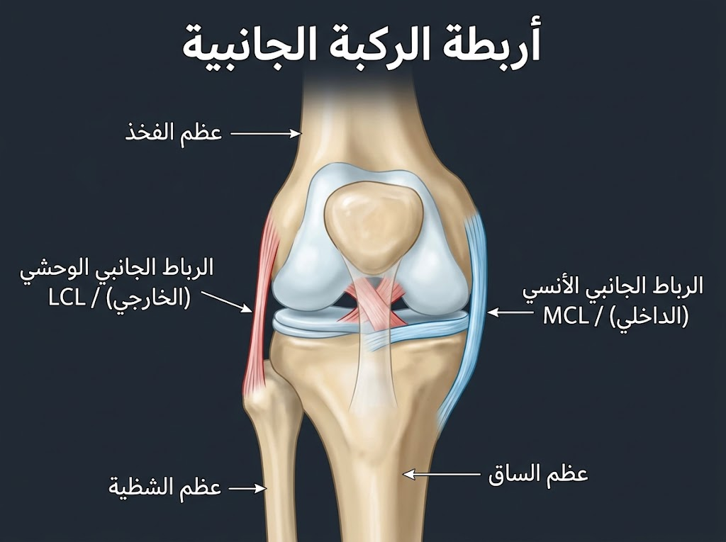
Key Differences Between the Two Ligaments:
| Feature | MCL (Medial) | LCL (Lateral) |
| Location | Inner side of the knee | Outer side of the knee |
| Mechanism of Injury | Direct blow to the outer side | Direct blow to the inner side or severe twisting |
| Prevalence | Most common — involved in 40% of sports knee injuries | Rarer — usually part of complex injuries |
| Primary Treatment | Conservative in most cases | Sometimes requires surgery |
Causes of Collateral Knee Ligament Tears
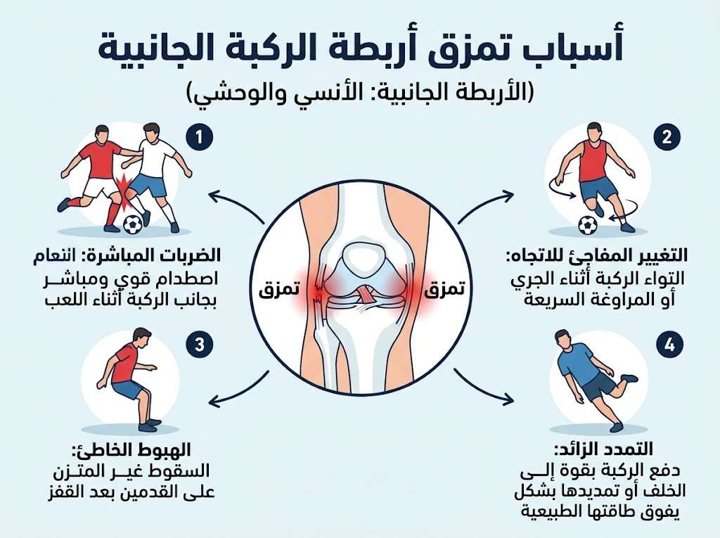
Collateral ligament tears usually result from a direct force that bends the knee inward or outward beyond its normal range of motion, or from a sudden twist during movement with the foot planted firmly on the ground.
- Direct blow to the outside of the knee — the leading cause of MCL injuries in soccer.
- Rapid twisting when changing direction (sports injuries).
- Falling with the knee twisted in a bent position — common in skiing and badminton.
- Car accidents (lateral impact force).
- Complex injuries: MCL + ACL + meniscus tear in the same incident.
Symptoms of Collateral Knee Ligament Tears
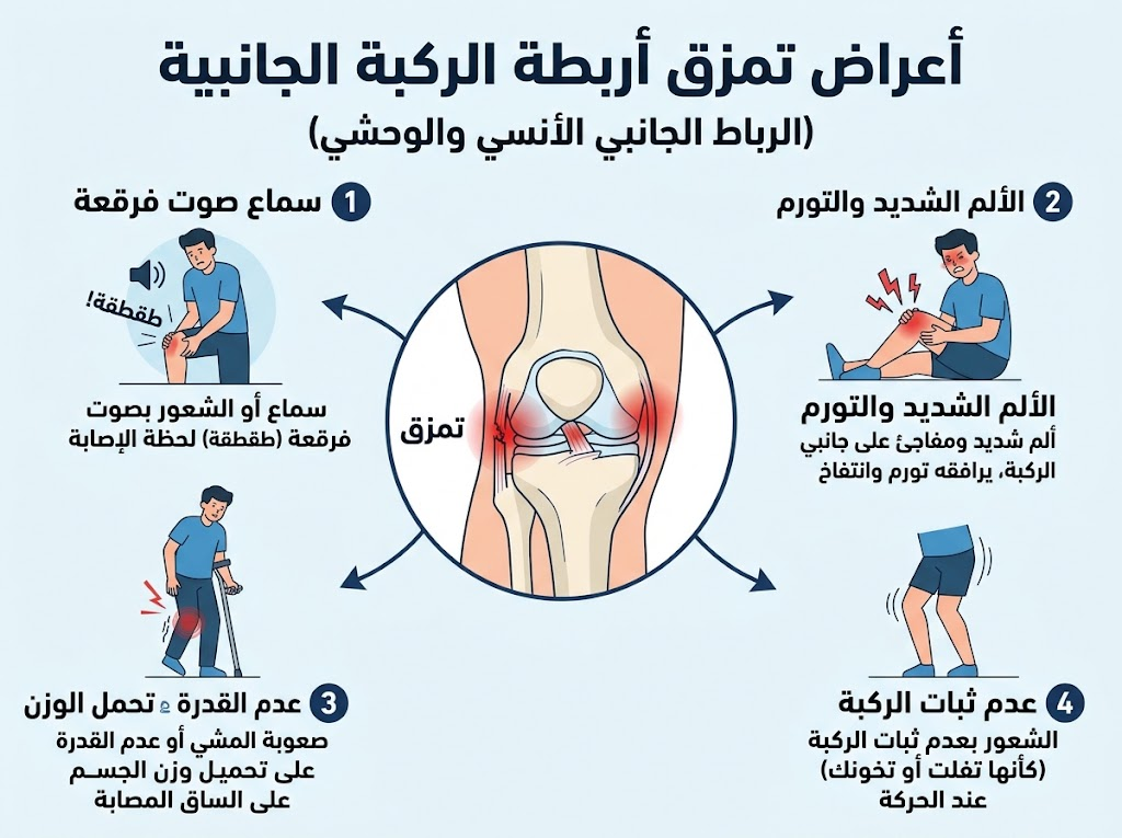
Symptoms of a collateral ligament tear include: immediate pain on the affected side of the knee, occasionally accompanied by a popping sound; localized swelling that appears within hours; a feeling of knee instability when standing or walking; and direct tenderness when pressing along the ligament’s path. Pain intensifies when the knee is tilted toward the side opposite the injury.
Signs to Differentiate the Grade of Injury:
- Audible popping sound at injury: Indicates a Grade II or III tear.
- Weight-bearing ability: Partially preserved in Grades I and II, lost in Grade III.
- Instability during Valgus/Varus Stress Test: Positive in Grades II and III.
- Diffuse swelling inside the joint: Suggests an associated injury (ACL or meniscus).
Grades of Collateral Knee Ligament Tears
| Grade | Tear Percentage | Pain | Stability | Recovery Time | Treatment |
| I — Sprain | < 25% of fibers | Mild to moderate | Normal | 2–4 weeks | Conservative |
| II — Partial | 25–75% | Moderate to severe | Slightly reduced | 4–8 weeks | Conservative + brace |
| III — Complete | > 75% (complete rupture) | Severe | Absent | 3–12 months | May require surgery |
How are knee ligament tears diagnosed?
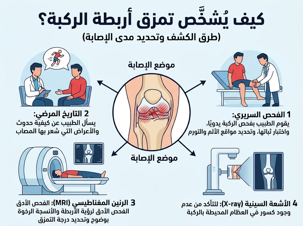
Diagnosis begins with a clinical examination (lateral stress testing of the knee at 0 and 30 degrees of flexion), followed by X-rays to rule out fractures, and an MRI to confirm the tear grade and detect any associated injuries.
- Valgus Stress Test: The knee is pushed inward to test the MCL.
- Varus Stress Test: The knee is pushed outward to test the LCL.
- Joint opening > 5 mm at 30 degrees of flexion: Indicates a partial or complete tear.
- MRI: Always necessary to rule out associated ACL or meniscus tears (which occur in 20–50% of severe MCL injuries).
Treatment of Knee Ligament Tears
Conservative Treatment (Grades I and II)
Grades I and II are treated conservatively in most cases, with a recovery rate exceeding 85%.
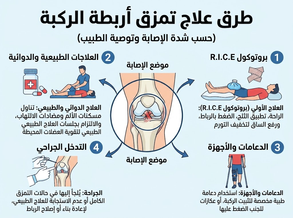
- PRICE protocol during the first 48–72 hours: Protection, Rest, Ice (20 minutes every 4 hours), Compression with an elastic bandage, and Elevation of the limb.
- Functional Brace: Stabilizes the knee and allows for a gradual return to motion in Grade II injuries.
- Physical Therapy: Strengthening the thigh and gluteal muscles, and proprioception (balance) exercises — starting after acute inflammation subsides.
- Crutches: Used to reduce weight-bearing during the first week for Grades II and III.
Medications for Knee Ligament Tears
| Category | Common Brand Names | Usage | Duration of Use |
| Oral NSAIDs | Ibuprofen, Diclofenac (Cataflam), Naproxen | Pain relief + anti-inflammatory | 5–10 days |
| Topical Anti-inflammatories | Voltaren Gel, Fastum Gel | Localized pain relief | 2 weeks |
| Muscle Relaxants | Methocarbamol (Robaxin), Thiocolchicoside | Relief of muscle spasms | 5–7 days |
| Proton Pump Inhibitors | Omeprazole | Stomach protection with NSAIDs | Throughout NSAID use |
Note: Oral corticosteroids are not routinely given for acute ligament injuries as they delay healing during the remodeling phase.
Injections for Knee Ligament Inflammation
In cases of chronic inflammation or persistent pain after 6 weeks of conservative treatment, ultrasound-guided local injections are administered directly into the affected ligament.
| Injection Type | Mechanism | Best Suited For | Expected Duration |
| Localized Cortisone | Rapid anti-inflammatory | Resistant acute inflammation | 4–8 weeks |
| PRP (Platelet-Rich Plasma) | Stimulates growth factors for repair | Chronic partial tears, sports rehab | 3–6 months |
| Prolotherapy (Dextrose) | Stimulates a localized healing response | Chronic ligament laxity | Repeated sessions |
Ultrasound guidance during injection is essential for precise delivery to the ligament; blind injections are not recommended.
Surgical Treatment (Grade III)
Surgery is required in cases of: complete LCL tears, complex injuries (e.g., MCL + ACL), or failure of conservative treatment after 3 months with persistent instability. Options include:
- Primary Repair: Possible within 3 weeks of injury before the ligament ends degrade.
- Reconstruction: Using a graft from the patient’s own tendon or a donor if intervention is delayed.
- Note: Most complete MCL tears heal without surgery due to the medial ligament’s excellent blood supply.
Recovery Period from Knee Ligament Tears
| Grade | Return to Normal Walking | Return to Light Sports | Full Return to the Field |
| I | Almost immediate | 1–2 weeks | 2–4 weeks |
| II | 3–7 days (with brace) | 3–6 weeks | 6–8 weeks |
| III (Conservative) | 2–3 weeks | 3–4 months | 4–6 months |
| III (Surgical) | 4–6 weeks | 4–5 months | 6–12 months |
Best Doctor for Knee Treatment in Cairo
Prof. Dr. Ibrahim Shaarawi, Professor of Orthopedic Surgery at Ain Shams University and Visiting Professor at Ohio State University, specializes in arthroscopic knee surgery and ligament reconstruction. He provides ultrasound-guided PRP and cortisone injections at his clinics in Nasr City, Fifth Settlement, and Mohandessin.

Frequently Asked Questions: Collateral Knee Ligament Tears
What is the anatomical difference between the MCL and LCL?
The two collateral ligaments stabilize the knee against side-to-side forces:
- Medial Collateral Ligament (MCL): A broad, flat band on the inside of the knee connecting the medial femoral condyle to the proximal tibia. It resists valgus (inward) stress and external rotation.
- Lateral Collateral Ligament (LCL): A cord-like extra-articular structure on the outside of the knee connecting the lateral femoral epicondyle to the fibular head. It resists varus (outward) stress and acts alongside the posterolateral corner (PLC).
How are collateral knee ligament tears clinically graded?
Severity is classified based on joint-line opening during physical stress testing:
- Grade 1 (Mild Sprain): Microscopic fiber tears, localized tenderness, and minimal swelling, with normal joint stability (<5 mm joint opening with a firm endpoint).
- Grade 2 (Partial Tear): Macroscopic disruption of fibers, significant local pain, localized ecchymosis, and moderate laxity (5–10 mm opening with a distinct endpoint).
- Grade 3 (Complete Tear): Full-thickness anatomical rupture with gross joint instability (>10 mm opening without a firm endpoint).
What are the hallmark symptoms of a collateral ligament injury?
Patients typically present with:
- Well-demarcated pain along the medial or lateral joint margin on palpation.
- Localized swelling or superficial bruising along the ligament path within 24 to 48 hours.
- Difficulty bearing weight or antalgic gait due to pain and guarding.
- Mechanical instability or subjective feeling of the knee “buckling” or “giving way” on uneven ground.
What injury mechanisms typically cause collateral ligament tears?
Collateral tears stem from high-demand sports contact or traumatic falls:
- MCL: Direct lateral contact forcing the knee inward (valgus stress), or non-contact planting and external tibial rotation (common in football, soccer, and skiing).
- LCL: Direct medial impact forcing the knee outward (varus stress), or hyperextension with varus loading, often occurring in motor vehicle accidents or direct tackles.
Can an MCL tear heal without surgical intervention?
Yes. The MCL has an extensive vascular network and an extra-articular envelope, providing superior biological healing capacity compared to intra-articular ligaments like the ACL. Over 90% of isolated Grade 1, 2, and 3 MCL injuries heal with conservative protocols utilizing protected range-of-motion bracing and progressive functional rehabilitation.
When is surgery indicated for collateral knee ligament tears?
Surgical management is indicated in specific, complex scenarios:
- Grade 3 LCL injuries involving the posterolateral corner (PLC), which carry poor natural healing potential.
- Multi-ligament injuries (e.g., knee dislocations or combined ACL/PCL/collateral disruption).
- Bony avulsion injuries (such as a Segond fracture or proximal fibular avulsion).
- Stener-like lesions where the distal torn end of the MCL displaces superficial to the pes anserinus tendons, blocking anatomical healing.
- Persistent rotatory or translational instability following completed conservative therapy.
How is a collateral ligament tear accurately diagnosed?
Comprehensive orthopedic evaluation involves:
- Clinical Stress Examination: Valgus stress testing (for MCL) and varus stress testing (for LCL) performed at both 0° (full extension) and 30° of knee flexion to assess isolated vs. combined ligament integrity.
- Magnetic Resonance Imaging (MRI): Gold standard for visualizing tear grade, anatomical location (femoral vs. tibial attachment vs. midsubstance), and concomitant meniscal or cruciate damage.
- Radiographs: Stress and standard AP/lateral views to rule out epiphyseal fractures, bony avulsions, or arthritic changes.
What is the expected recovery timeline for collateral ligament tears?
Rehabilitation timelines scale with injury grade:
- Grade 1: 1 to 3 weeks for full, pain-free athletic return.
- Grade 2: 4 to 6 weeks with a hinged brace, progressive strengthening, and sports-specific functional drills.
- Grade 3 (Non-Operative): 8 to 12 weeks of structured bracing, isometric loading, and proprioceptive conditioning.
- Surgical Reconstruction: 6 to 9 months, adhering to strict graft maturation and functional return-to-sport metrics.
What immediate first-aid steps should be taken after a collateral ligament injury?
Immediate management follows the PRICE protocol within the first 48 hours:
- Protect: Immobilize the knee with a brace or knee immobilizer to prevent further coronal displacement.
- Rest: Avoid direct weight-bearing; utilize axillary crutches until clinical assessment.
- Ice: Apply cryotherapy for 15–20 minutes every 2 hours to diminish metabolic demand and edema.
- Compress: Apply an elastic bandage evenly from distal to proximal to control effusion.
- Elevate: Elevate the limb above heart level to facilitate lymphatic drainage.
What is the clinical role of hinged bracing and physical therapy in recovery?
A hinged knee brace shields repairing ligamentous scar tissue by offloading coronal (varus/valgus) stresses while preserving functional sagittal flexion and extension. Concurrently, physical therapy maintains quadriceps and hamstring tone, restores normal gait mechanics, prevents arthrofibrosis, and trains neuromuscular sensorimotor feedback for joint protection.
REFERENCES
- American Academy of Orthopaedic Surgeons (AAOS), OrthoInfo: Collateral Ligament Injuries
- American Academy of Orthopaedic Surgeons (AAOS), OrthoInfo: Combined Knee Ligament Injuries
- https://my.clevelandclinic.org/health/diseases/21710-lcl-tears
(Cleveland Clinic — LCL tear: symptoms, 3 grades, surgical & non-surgical treatment)
https://my.clevelandclinic.org/health/diseases/21979-mcl-tear
(Cleveland Clinic — MCL tear: Grade 1 (<10% fibers), Grade 2 (partial), Grade 3 (complete) — recovery times)
https://orthoinfo.aaos.org/en/diseases–conditions/chronic-shoulder-instability/
(AAOS — ligament injury classification)
https://www.sports-injury-physio.com/post/lateral-collateral-ligament-lcl-tears-or-sprains-symptoms-treatment-recovery-times
(LCL tear — MRI grading, recovery times by grade: Grade 1 = 4–6 weeks, Grade 2 = 8–12 weeks, Grade 3 = bracing 2 weeks — updated 2026)
https://www.sportsinjuryclinic.net/acute-knee-injuries/lateral-knee-ligament-sprain
(LCL sprain — symptoms, 3 grades, varus stress test, massage, treatment — updated April 2026)
https://corycalendinemd.com/lcl-tear-symptoms-treatment-recovery/
(LCL tear guide — 3 grades, healing time 3–12 weeks, when surgery is needed — Oct 2026)
https://regenexx.com/condition/knee/lateral-collateral-ligament-tear/
(LCL tear — symptoms: popping, instability, can’t straighten knee — conservative vs surgical)
https://www.ncbi.nlm.nih.gov/pmc/articles/PMC9275808/
(MCL/LCL injuries with ACL — 77% association rate — diagnosis by MRI and arthroscopy)