
Anatomy of the knee joint with pictures — its bones, ligaments, cartilages, and normal appearance on X-rays, along with the most common knee injuries, causes of pain, and treatment in Cairo.
What is the knee joint?
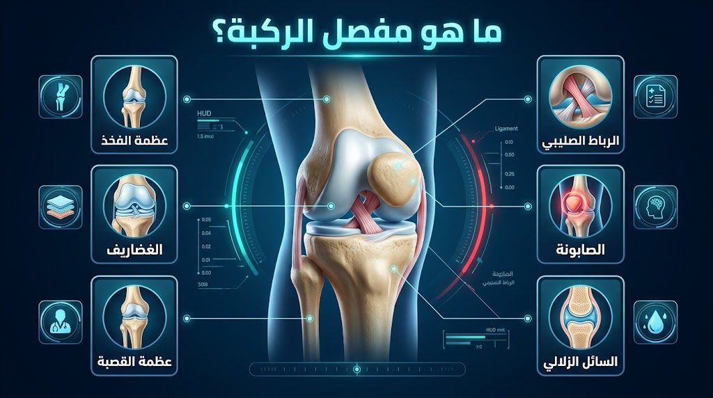
The knee joint is the largest joint in the human body. It is a modified hinge joint connecting the femur (thigh bone) to the tibia (shin bone) and the patella (kneecap). It allows for flexion and extension between 0 and 135 degrees, with limited internal and external rotation. Its stability relies primarily on the four ligaments and surrounding muscles, rather than the shape of the bones.
Components of the Knee Joint with Pictures
Bones forming the knee
Three bones make up the knee joint:
- Femur (thigh bone): The lower end forms into two condyles that make up the upper joint surface.
- Tibia (shin bone): The upper end bears the tibial plateau as the lower articular surface.
- Patella (kneecap): A sesamoid bone that glides in the femoral groove, multiplying the force of the anterior thigh muscle (quadriceps) during bending.
- Note: The Fibula (calf bone) does not participate in forming the knee joint despite its close proximity to it.
The Four Main Ligaments
| Ligament | Location | Function | Most susceptible to |
| ACL (Anterior Cruciate Ligament) | Inside the joint | Prevents the tibia from sliding forward | Sports twisting injuries |
| PCL (Posterior Cruciate Ligament) | Inside the joint | Prevents the tibia from sliding backward | Car accidents |
| MCL (Medial Collateral Ligament) | Outside the joint | Prevents outward deviation | Blows to the outer side |
| LCL (Lateral Collateral Ligament) | Outside the joint | Prevents inward deviation | Rare — complex injuries |
Cartilages and Synovial Bursae
- Meniscus: Two cartilaginous discs, medial and lateral, acting as shock absorbers and improving load distribution — their tearing is one of the most common knee injuries.
- Articular Cartilage: A smooth layer 2–4 mm thick lining the bone surfaces, which wears away in osteoarthritis.
- Synovial Membrane: Produces synovial fluid that nourishes the cartilage and lubricates the joint.
- Bursae: 14 synovial sacs around the knee, the most famous being the Prepatellar Bursa, which becomes inflamed from prolonged kneeling.
Normal Appearance of the Knee Joint on X-rays
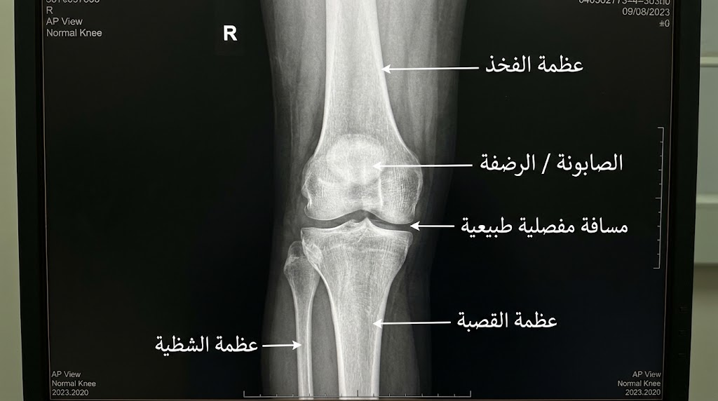
In a normal standing X-ray (AP View), the joint space between the femur and tibia is 3–5 mm, the limb axis tilts 5–7 degrees outward (Valgus), and there are no bony spurs (osteophytes) or space narrowing. A decrease below 2 mm indicates advanced osteoarthritis.
Normal X-ray Criteria:
| Radiological Criterion | Normal Value | Indication of Deviation |
| Joint Space (AP) | 3–5 mm | < 2 mm = advanced osteoarthritis |
| Knee Axis (Femorotibial angle) | 5–7° Valgus | > 10° = deformity |
| Insall-Salvati ratio (Patella) | 0.8–1.2 | > 1.2 = high riding patella (Patella Alta) |
| Bone Density | Homogeneous | Radiolucent spots = necrosis or cyst |
CT scans are used to evaluate fractures and axes. MRI is the gold standard for diagnosing ligament and cartilage injuries.
Most Common Knee Injuries
| Injury | Most Affected Category | Defining Sign | Treatment Option |
| ACL Tear | Youth and sports injuries | Popping sound + inability to bear weight | Arthroscopic reconstruction |
| Meniscus Tear | All ages | Joint locking + pain when twisting | Arthroscopic repair/excision |
| Knee Osteoarthritis (OA) | > 50 years old, overweight | Gradual pain + morning stiffness | Injections + physical therapy ← Replacement |
| Patellar Dislocation | Teenage girls | Sensation of the kneecap slipping out | Conservative treatment ← Surgical fixation |
| Patellar Tendinitis (Jumper’s Knee) | Jumping athletes | Pain directly below the kneecap | Rest + PRP |
| MCL Tear | Physical contact in sports | Inner side pain + instability | Mostly conservative |
| Terrible Triad | Violent accidents | ACL + MCL + meniscus together | Complex arthroscopic surgery |
Causes of Knee Pain
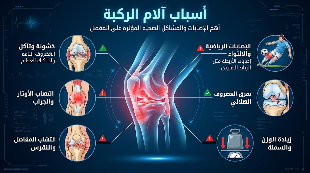
Knee pain is not a single disease but a symptom of multiple causes. The primary distinction: pain that increases with movement and subsides with rest indicates osteoarthritis, whereas continuous night pain requires ruling out a tumor, and sudden acute pain after an injury means an immediate MRI is needed.
Classification of Causes:
- Degenerative: Knee osteoarthritis (most common in Egypt after age 50).
- Traumatic: Ligament and cartilage tears, fractures.
- Inflammatory: Rheumatoid arthritis, gout.
- Mechanical: Patellofemoral osteoarthritis, Iliotibial band syndrome.
- Referred from the hip: Hip pain is sometimes referred to the knee — especially in children (often misdiagnosed).
- Tumors: Rare, but night pain is a warning sign not to be ignored.
Treatment of Knee Pain
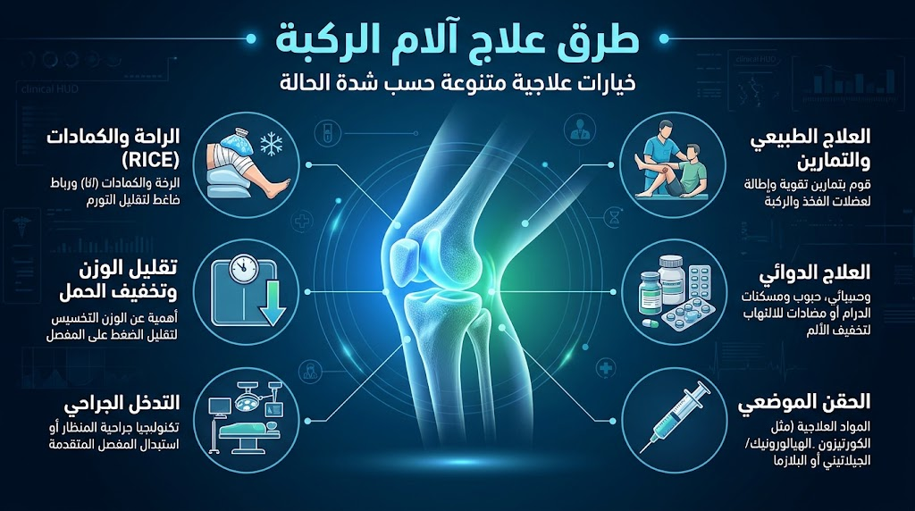
The treatment option is determined by the cause and its severity, not the pain alone. Mild osteoarthritis is treated with exercises and weight loss before any medication. An acute sports injury requires radiological diagnosis before starting any protocol.
- Conservative: Relative rest, icing for 20 minutes 4–6 times daily in the first 48 hours, NSAIDs (painkillers).
- Physical Therapy: Strengthening surrounding muscles, improving balance, TENS for pain relief.
- Knee Injections: Cortisone (acute inflammation) → Hyaluronic Acid (Grade I–III OA) → PRP (mild OA, sports injuries).
- Arthroscopic Surgery: ACL reconstruction, meniscus repair, joint debridement.
- Knee Joint Replacement: For Grade IV osteoarthritis after all the above have failed.
Best Doctor for Knee Treatment in Cairo
Prof. Dr. Ibrahim Shaarawi, Professor of Orthopedic Surgery at Ain Shams University and Visiting Professor at Ohio State University, diagnoses and treats all knee injuries from meniscal and ligament arthroscopy to total joint replacement. He has clinics in Nasr City, Fifth Settlement, and Mohandessin.

Frequently Asked Questions: Anatomy & Function of the Knee Joint
What bones form the human knee joint?
The knee joint is composed of three articulating bones forming two interconnected functional compartments:
- Femur (Thigh Bone): Features two rounded distal condyles that interface with the tibial plateau.
- Tibia (Shin Bone): Provides the flat, weight-bearing tibial plateau that supports bodily loads.
- Patella (Kneecap): A sesamoid bone embedded in the quadriceps/patellar tendon that glides within the femoral trochlear groove.
The fibula runs parallel to the lateral tibia but does not form part of the true weight-bearing knee joint articulation.
What type of joint is the knee anatomically?
The knee is classified as a modified hinge joint (or bicondylar synovial joint). While primary motion occurs in the sagittal plane via flexion and extension (roughly 0° to 140°), asymmetry between the medial and lateral femoral condyles allows secondary rotational translation, permitting internal and external rotation when the knee is flexed.
What are the four primary stabilizing ligaments of the knee joint?
Four primary ligaments maintain passive kinematic stability:
- Anterior Cruciate Ligament (ACL): Traverses obliquely within the intercondylar notch to prevent anterior tibial translation and control rotatory subluxation.
- Posterior Cruciate Ligament (PCL): The strongest intra-articular stabilizer, preventing posterior translation of the tibia on the femur.
- Medial Collateral Ligament (MCL): A broad, extra-articular band shielding the medial compartment from valgus (knock-knee) collapse.
- Lateral Collateral Ligament (LCL): A discrete cord-like stabilizer resisting varus (bow-leg) opening and posterolateral rotational forces.
What is the biological function of the knee menisci?
The medial and lateral menisci are wedge-shaped fibrocartilaginous structures situated on the tibial plateau. Key functions include:
- Load Transmission: Bearing 50% to 70% of overall compressive contact loads across the knee.
- Shock Absorption: Dampening ground reaction impacts during heel strike, running, and jumping.
- Congruence & Stability: Converting flat tibial surfaces into concave sockets matching the rounded femoral condyles.
- Lubrication: Spreading synovial fluid evenly over articular hyaline cartilage surfaces.
What is articular cartilage, and why is its damage difficult to heal?
Articular (hyaline) cartilage is a dense, glass-like matrix composed of type II collagen, proteoglycans, and water, providing a frictionless gliding interface. Unlike other vascular tissues, mature articular cartilage is aneural and avascular. Without direct blood delivery, chondrocytes cannot mount a standard inflammatory repair response, making mechanical wear, chondral fissures, and full-thickness defects permanent without surgical or regenerative intervention.
What is the function of synovial fluid and the synovial membrane in the knee?
The knee is enclosed in an extensive fibrous capsule lined internally by the synovial membrane. This membrane filters blood plasma and synthesizes hyaluronic acid and lubricin, producing clear synovial fluid. The fluid performs non-Newtonian hydrodynamic lubrication (becoming thinner under rapid shearing forces) while delivering glucose, oxygen, and amino acids to avascular articular cartilage via passive diffusion.
What is the ‘screw-home mechanism’ of the knee joint?
The screw-home mechanism is an involuntary rotation that locks the knee joint during the final 10° of terminal extension. Due to the larger articular surface area of the medial femoral condyle relative to the lateral, the tibia must rotate externally roughly 5° to 10° relative to the femur. This tight-packs the cruciate ligaments and condyles into maximal congruency, allowing prolonged upright standing with minimal active quadriceps energy consumption. The deep popliteus muscle contracts to internally rotate the tibia, “unlocking” the knee to permit subsequent flexion.
What are the primary muscle groups that move and dynamically stabilize the knee?
Dynamic movement and secondary joint stabilization rely on coordinated muscular contraction:
- Quadriceps Femoris: Extends the knee; controls eccentric loading (such as descending stairs) and regulates patellar tracking.
- Hamstrings: Flexes the knee and acts as an essential functional synergy with the ACL by resisting anterior tibial displacement.
- Popliteus: Acts as the primary rotator to unlock the knee from terminal extension and protects the lateral meniscus.
- Gastrocnemius: Crosses the posterior joint to assist knee flexion and reinforce posterior capsule stability.
What causes a joint effusion (‘water on the knee’)?
Intra-articular effusion (swelling within the capsule) results from an inflammatory or traumatic response of the synovial membrane. Rapid-onset swelling within 1 to 2 hours of trauma typically indicates hemarthrosis (bloody effusion, commonly from an acute ACL tear or osteochondral fracture). Delayed swelling appearing 12 to 24 hours later usually signifies reactive synovitis driven by meniscal tears, chondral abrasion, osteoarthritis flares, or systemic inflammatory arthritides.
What are the common degenerative and traumatic conditions affecting the knee?
Frequent clinical presentations encompass:
- Osteoarthritis: Progressive degeneration of articular cartilage and subchondral bone remodeling.
- Ligament Disruptions: Non-contact rotational tears (ACL) and contact-induced stress ruptures (MCL/PCL/LCL).
- Meniscal Tears: Acute radial, longitudinal, or bucket-handle tears in young athletes; horizontal cleavage tears in older populations.
- Extensor Mechanism Disorders: Patellofemoral pain syndrome, patellar dislocation, and jumper’s knee (patellar tendinopathy).
- Cysts & Bursitis: Prepatellar bursitis (housemaid’s knee) and popliteal (Baker’s) cysts secondary to chronic effusion.