
Shoulder osteoarthritis (glenohumeral osteoarthritis) is a progressive condition characterized by the gradual erosion of the cartilage lining the shoulder joint surfaces. This degradation results in direct bone-on-bone friction, leading to increasing pain and stiffness over time. While it most commonly affects individuals over the age of 60, it may occur earlier following past trauma, fractures, or recurrent dislocations. Treatment typically begins conservatively, escalating in intensity based on symptom severity.
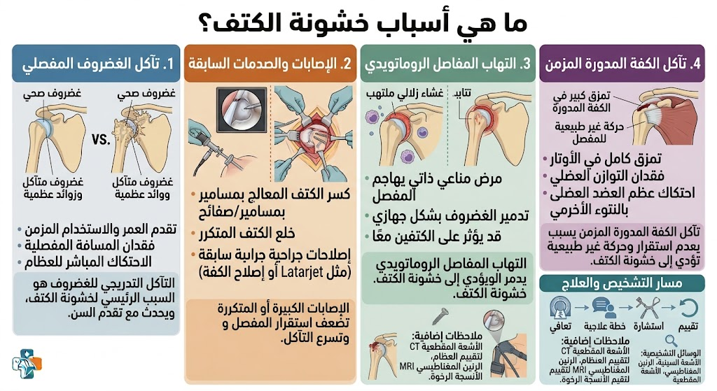
Causes of Shoulder Osteoarthritis
- Aging: The most common cause; cartilage naturally wears down over decades.
- Post-traumatic Injury: Previous fractures of the humeral head or glenoid cavity accelerate cartilage erosion.
- Chronic Rotator Cuff Tears: Untreated long-term tears alter joint mechanics, causing uneven wear (rotator cuff arthropathy).
- Recurrent Dislocation: Each episode damages the cartilage, predisposing the joint to early-onset osteoarthritis.
- Chronic Inflammatory Conditions: Diseases like rheumatoid arthritis or gout directly attack joint cartilage.
- Avascular Necrosis: The death of bone tissue in the humeral head due to compromised blood supply.
Symptoms of Shoulder Osteoarthritis
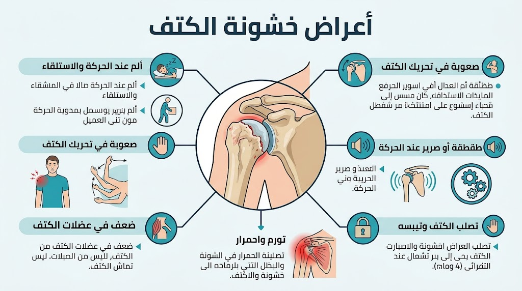
Symptoms typically evolve gradually:
- Deep Joint Pain: Intensifies with activity and subsides with rest.
- Stiffness: Limits the range of motion (ROM), particularly regarding rotation and overhead lifting.
- Crepitus: Audible clicking or grinding sounds during movement.
- Night Pain: Becomes more pronounced as the condition progresses. In advanced stages, pain may radiate toward the neck and arm.
Osteoarthritis Grading
| Grade | Description | Symptoms |
| Grade 1 | Minor cartilage erosion | Mild pain after exertion; relieved by rest. |
| Grade 2 | Moderate erosion with joint space narrowing | Regular pain with morning stiffness. |
| Grade 3 | Significant erosion with bone spurs | Persistent pain; stiffness hinders daily activities. |
| Grade 4 | Near-total cartilage loss | Severe pain; significant functional disability. |

Treatment Modalities
Conservative Management (Grades 1–2)
- Pharmacology: NSAIDs (e.g., ibuprofen, diclofenac) mitigate pain and inflammation. Use should be intermittent to avoid gastrointestinal side effects.
- Physical Therapy (PT): The cornerstone of management. Dedicated daily exercises focus on ROM and strengthening the rotator cuff.
- Activity Modification: Avoid movements that exacerbate pain, heavy overhead lifting, and repetitive stress.
- Intra-articular Injections: Corticosteroids, hyaluronic acid, or Platelet-Rich Plasma (PRP) can be effective.
- Heat Therapy: 15–20 minutes of warm compresses can alleviate morning stiffness.
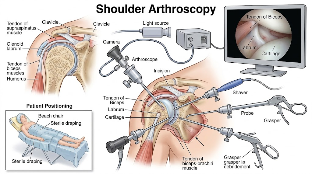
Arthroscopic Intervention (Grades 2–3)
Arthroscopy is used to clean the joint (debridement) and remove bone spurs or inflamed tissue. While it provides temporary symptom relief (1–3 years), it cannot replace lost cartilage.
Joint Replacement (Grades 3–4)
When conservative measures fail, total shoulder arthroplasty offers a long-term solution with a success rate exceeding 90%.
- Total Shoulder Replacement (Anatomical): Used when the rotator cuff remains intact.
- Reverse Shoulder Replacement: Used for severe arthritis combined with massive rotator cuff tears; the design relies on the deltoid muscle rather than the damaged cuff.

Frequently Asked Questions: Shoulder Osteoarthritis
1. What is shoulder osteoarthritis (glenohumeral osteoarthritis)?
Shoulder osteoarthritis involves the progressive degeneration of the smooth articular cartilage lining the humeral head (ball) and the glenoid cavity (socket). As the cartilage wears down, friction between exposed bone surfaces results in mechanical pain, secondary joint inflammation, and peripheral osteophyte (bone spur) formation.
2. What are the primary causes and risk factors for shoulder arthritis?
Primary shoulder osteoarthritis is commonly associated with aging and genetic predisposition. Secondary causes include post-traumatic damage (prior fractures or recurrent dislocations), chronic massive rotator cuff tears leading to cuff tear arthropathy, avascular necrosis (AVN) of the humeral head, and systemic inflammatory arthritis such as rheumatoid arthritis.
3. What are the hallmark symptoms of shoulder osteoarthritis?
The most frequent symptoms include deep, aching joint pain exacerbated by activity, prominent night pain that disturbs sleep when lying on the affected side, progressive stiffness with restricted range of motion (especially external rotation and overhead elevation), and palpable or audible crepitus (grinding/clicking sensations).
4. How does shoulder osteoarthritis differ from rotator cuff tendinopathy or impingement?
In rotator cuff tendinopathy or impingement, pain typically occurs during specific active arcs of motion, while passive range of motion (when the clinician moves the arm) is largely preserved. In glenohumeral osteoarthritis, both active and passive motion are progressively restricted due to mechanical osteophytes and capsular contracture.
5. How is shoulder osteoarthritis diagnosed?
Diagnosis begins with a focused clinical history and physical examination. Standard plain radiographs (true AP/Grashey, axillary, and scapular Y views) remain the gold standard, demonstrating joint space narrowing, subchondral sclerosis, cyst formation, and inferior humeral osteophytes. CT scans are utilized for surgical planning to evaluate glenoid bone loss and version, while MRI evaluates the integrity of the rotator cuff muscles and tendons.
6. What non-surgical treatments are effective for managing symptoms?
First-line conservative strategies include activity modification (avoiding repetitive overhead loading), targeted physical therapy focusing on capsular stretching and periscapular stabilization, non-steroidal anti-inflammatory drugs (NSAIDs) or acetaminophen, thermal modalities (ice/heat), and intra-articular injections.
7. Do joint injections cure shoulder osteoarthritis?
Intra-articular injections (such as corticosteroids, hyaluronic acid/viscosupplementation, or platelet-rich plasma [PRP]) do not reverse cartilage loss or restore worn cartilage. Instead, they provide targeted symptomatic pain relief and decrease synovitis, opening a therapeutic window for physical rehabilitation and improving daily function.
8. When is surgery or shoulder replacement recommended?
Surgical intervention is indicated when conservative measures have failed to control persistent, severe pain (particularly intractable nocturnal pain), and when structural joint destruction significantly impairs basic activities of daily living.
9. What is the difference between Anatomic Total Shoulder Arthroplasty (TSA) and Reverse Total Shoulder Arthroplasty (rTSA)?
Anatomic Total Shoulder Arthroplasty (TSA) replicates native anatomy by replacing the ball with a metal prosthetic head and the socket with a plastic glenoid component; it requires a functioning, intact rotator cuff. Reverse Total Shoulder Arthroplasty (rTSA) reverses the ball and socket configuration (placing a spherical glenosphere on the socket and a concave cup on the humerus); this design allows the deltoid muscle to lift the arm when the rotator cuff is deficient, torn, or severely compromised.
10. What lifestyle and ergonomic modifications help protect an arthritic shoulder?
Patients are advised to keep objects close to the body when lifting, avoid sudden heavy ballistic loading or repetitive overhead strain, maintain daily low-impact range-of-motion exercises (such as pendulum or pulley stretches), optimize sleeping posture using supporting pillows, and maintain good thoracic and cervical spine alignment.
References
- American Academy of Orthopaedic Surgeons (AAOS), OrthoInfo: Arthritis of the Shoulder
- American Academy of Orthopaedic Surgeons (AAOS), OrthoInfo: Shoulder Joint Replacement